Download
1 / 73
760 likes | 1.05k Vues
The Senses. Anatomy & Physiology I Chapter 11. The Senses. Protect a person by detecting changes in the environment Stimulus – an environmental change that initiates or stimulate a nerve impulse

E N D
The Senses Anatomy & Physiology I Chapter 11
The Senses • Protect a person by detecting changes in the environment • Stimulus – an environmental change that initiates or stimulate a nerve impulse • Sensation – when a specialized area of the cerebral cortex interprets the nerve impulse and the stimulus then becomes something we experience • Environmental change >> stimulus >>threshold stimulus >> sensory receptors >> sensory neuron>> CNS >> interpretation>> sensation (what we experience or sense) • Threshold stimulus - minimum amount of stimulus required to generate a nerve impulse
Sensory Receptors – Types based on Structure • Free dendrite of a sensory neuron; Example, pain, temperature • End-organ - the modified dendrite ending of a sensory neuron enclosed in a capsule -Pacinian & Meissner corpuscles for pressure & touch receptors • Specialized cell associated with an afferent neuron (afferent/ascending/to the CNS); rods & cones in the eye
Sensory Receptors – Types based on Stimulus • Chemoreceptors – detect chemicals in solution; taste & smell • Photoreceptors – respond to light; eye retina • Thermoreceptors – detect temperature change; skin • Mechanoreceptors – respond to movement, vibration, pressure, stretch; located in skin, joints (monitor body position), hearing & equilibrium in ear
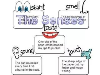
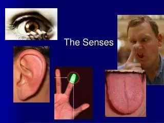
Types of senses based on the distribution of their receptors • Special senses; sensory receptors localizedin a special sense organ • Vision • Hearing • Equilibrium • Taste • Smell • General senses; sensory receptors widely distributed throughout the body • Pressure • Temperature • Pain • Touch • Position
The Eye - Protective Structures • Skull bones form walls of eye orbit; over half of posterior eye • Upper & lower eyelids; anterior eye • Eyelashes & eyebrows; anterior eye • Conjunctiva; lines inner surface of eyelids & covers the visible portion of the white of the eye (sclera); produces mucus & is highly vascular • Lacrimal glands; produce tears which lubricate & produce enzyme that protects against infection • Flow from superior lateral eye into inferior medial nasolacrimal duct
Coats of the Eyeball • 3 tunics (coats) • Sclera – outermost tunic made of tough connective tissue; white of the eye because made of collagen & no blood vessels • Choroid – coat made of delicate connective tissue; extensive blood supply which are visualized during eye exam; prevents light from scattering throughout eye • Retina – actual receptor layer; contains rods & cones which generate visual nerve impulses
What are some structures that protect the eye? • What are the names of the tunics of the eyeball?
Pathway of Light Rays & Refraction • Refraction – bending of light rays as they pass from one substance to another substance of a different density • Allows light from a very large area to be focused on a very small area of the retina • Cornea • Aqueous humor • Lens • Vitreous body
Light Refraction in the Eye • Cornea – transparent, colorless continuation of sclera that covers the pupil; the window of the eye • Aqueous humor – watery fluid that fills the eye anterior to the lens; aids in refraction & maintains eye shape • Lens – clear circular structure with biconcave surface made of firm, elastic material; can change in thickness & focus near or far • Vitreous body – soft gel that fills entire space posterior to the lens; aids in refraction & maintains eye shape
Layers of the Retina • Pigmented layer – deepest layer just anterior to choroid • Rods & cones – receptors of the eye • Connecting neurons that carry impulses toward the optic nerve
Rods • Rods – highly sensitive to light • function in dim light but do not provide sharp image • Dark adaptation; the time it takes for rods to begin working in a darkened area • 120 million each retina • Distributed towards the periphery of the retina • See shades of gray; no colors • Rhodopsin – visual purple pigment that is sensitive to light; requires vitamin A; lack of this pigment leads to night blindness
Cones • Cones – sensitive to color • Function only in bright light • 6 million per retina • Localized in center of retina • fovea centralis, pit near the optic nerve; area of greatest visual acuity • surrounded by the macula lutea • Optic disk; point where the optic nerve arises in the retina; no rods or cones in this area; blind spot on the retina • Sees sharp images • Pigments sensitive to red, green, blue • Hereditary lack of pigment can lead to colorblindness in males
Fovea & Macula lutea Fovea (dark pink) & Macula lutea (yellowish)
Visual Impulses • Light stimulates rods & cones which stimulate neurons that eventually merge to form the optic nerve (CN II) • Some optic nerve fibers crosses at optic chiasma • Visual center in the occipital cortex of the cerebrum interprets
Eye Muscles – Extrinsic & Intrinsic Groups • Extrinsic • 6 voluntary on outer surface of eye • Controlled by CN III, IV, VI (oculomotor, trochlear, abucens) • Convergence – pulling the eyeballs in a coordinated fashion so there is one visual field
Eye Muscles – Extrinsic & Intrinsic Groups • Intrinsic • Involuntary muscles within the eyeball • Controlled by CN III (oculomotor) • Iris; pigmented part of the eye composed of 2 sets of muscles that control pupil size • Circular muscle constricts in bright light • Radial muscle constricts in dim light • Ciliary muscle; holds the lens of the eye in place by suspensory ligaments • Accomodation – ciliary muscle constriction changes the shape of the lens to allow for near & far vision
The light rays from a close object diverge (separate) more than do the light rays from a distant object >>the lens must become more rounded to bend the light rays more • When the ciliary muscle is relaxed, tension on the suspensory ligaments keeps the lens in a more flattened shape. For close vision, the ciliary muscle contracts • For close vision, the ciliary muscle contracts, which draws the ciliary ring forward and relaxes tension on the suspensory ligaments. The elastic lens then recoils and becomes thicker
Nerve Supply to Eye Two sensory nerves supply the eye: • CNII – optic nerve; carries visual impulses from retinal rods & cones to the thalamus (diencephalon) to the visual center in occipital lobe of cerebrum • CNV – trigeminal nerve, opthalmic branch; carries pain, touch & temperature impulses from the eye to the brain • There are no retinal rods and cones in the area of the optic nerve. Consequently, no image can form on the retina at this point, which is known as the blind spot or optic disk
Nerve Supply to the Eye Three nerves carry motor impulses to the eyeball muscles: • CNIII – oculomotor; largest motor nerve to the eyeball; supplies voluntary & involuntary muscles to all but two eye muscles • CNIV – trochlear; supplies superior extrinsic eye muscle • CNVI – abducens; supplies lateral extrinsic eye muscles
Steps in Vision • Light refracts (bends) • Muscles of the iris adjust the pupil • Ciliary muscle adjusts the lens (accomodation) • Extrinsic eye muscles produce convergence (coordinate to allow one visual field) • Light stimulates rods & cones • Optic nerve transmits impulses to thalamus • Thalamus transmits impulses to occipital lobe • Occipital lobe cortex interprets impulses
Errors of Refraction • Hyperopia – farsightedness • Usually due to abnormally short eyeball (flat cornea) • Light focuses behind retina • The lens can thicken only to a given limit to accommodate for near vision • move an object away from the eye to see it clearly • Glasses with convex lenses that increase light refraction • Myopia – nearsightedness • Usually due to abnormally long eyeball • Light focuses in front of retina • Distant objects appear blurred and may appear clear only if brought near the eye • A concave lens corrects for myopia • Astigmatism – blurred vision • Cornea or lens curves irregularly, bending light incorrectly
A. Normal B. MyopiaC. Hyperopia D. Astigmatism
Eye Disorders • Strabismus – deviation of the eye due to lack of coordination of eye muscles • In convergent strabismus, the eye deviates toward the nasal side • In divergent strabismus, the affected eye deviates laterally. • if not corrected brain will not develop to see properly • Amblyopia – loss of vision in a healthy eye because it cannot work properly with the other eye
Eye Disorders • Conjuntivitis – inflammation of conjunctiva • Pinkeye – conjunctivitis caused by infection; is usually caused by cocci or bacilli • Inclusion conjunctivitis is an acute eye infection caused by Chlamydia (AKA Trachoma) • Corneal laceration – most common eye injury & if untreated can result in blindness • Cornea is avascular so it is possible to receive transplant without rejection • Enucleation – removes the eye due to traumatic injury • Cataract – opacity of the lens which can lead to blindness • Glaucoma – excess pressure in the eyeball due to aqueous humor not being reabsorbed into blood
Disorders related to retina • Diabetic retinopathy – retina damaged by vascular hemorrhages & overgrowth • Retinal detachment – retina separates from underlying layer as the result of trauma or fluid accumulation between tunics of eye • Macular degeneration – macula lutea deteriorates & distorts visual field
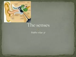
The Ear - Structure • Outer ear – external ear to the tympanic membrane • Middle ear – contains 3 bones (ossicles) of ear & eustachian tube • Inner ear – contains sensory receptors for hearing & equilibrium
Outer Ear • Pinna – aka auricle; the external ear • External auditory canal – aka external auditory meatus • Contains ceruminous glands • Tympanic membrane – aka eardrum • Vibrates as sound waves enter the ear
Middle Ear - Ossicles • Small cavity containing 3 ossicles (bones) that amplify sound waves received by tympanic membrane • Malleus (hammer) – attached to tympanic membrane by handle & head attaches to incus • Incus (anvil) – connects to malleus & stapes • Stapes (stirrup) – connects to oval window, the membrane of inner ear
Middle Ear – Eustachian Tube • Eustachian tube – connects middle ear to pharynx • Allows pressure to equalize on the 2 sides of tympanic membrane
Inner Ear aka Bony Labyrinth • 3 separate divisions of sensory receptors • Vestibule, semicircular canals & cochlea • Perilymph – fluid of inner ear • Membranous labyrinth – within bony labyrinth & filled with endolymph
Inner Ear aka Bony Labyrinth • Vestibule – 2 bony chambers that contain equilibrium receptors • Semicircular canals – 2 bony tubes that contain equilibrium receptors
Inner Ear aka Bony Labyrinth • Cochlea – bony coil that contains hearing receptors • Round window – membrane through which sound waves leave the inner ear
Hearing • Organ of Corti – sensory organ of hearing • Consists of ciliated receptor cells • Located inside membranous cochlea aka cochlear duct • Wave against the roof of the cochlear duct (tectorial membrane) • Stimulates cochlear nerve (auditory branch of vestibulocochlear nerve CN VIII) • Sound waves leave inner ear through round window
Steps of Hearing • Sound waves vibrate tympanic membrane • Amplified by ossicles in middle ear • Reach the oval window & create waves in the inner ear fluids • Vibrating the chochlear duct • Causing cilia or organ of Corti to wave against the tectorial membrane • Cochlear nerve stimulated (branch of CNVIII) • The temporal lobe of cerebrum interprets stimuli
Hearing • Organ of Corti differentiates both pitch (tone) & intensity (loudness) • Higher pitched tones near the base • Lower pitched tones near the top • Loud sounds stimulate more cells & produce more vibrations, sending more impulses to the brain
Equilibrium - Balance • Sensory receptors are ciliated cells located in vestibule & semicircular canals • Shifting in position of cilia within the thick fluid that surrounds them generates a nerve impulse • Send impulses to vestibular branch of CN VIII (vestibulocochlear)
Equilibrium - Balance Detail of Macula • Static equilibrium- Sensing the position of head or body when moving in a straight line • Due to vestibule receptors known as macula • Fluid surrounding maculae contains crystals called otoliths which drag the fluid & increase pull of gravity