The Senses
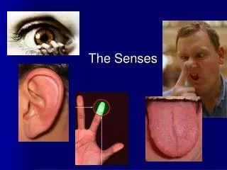
The Senses. The Senses. General senses of touch Temperature Pressure Pain Special senses Smell Taste Sight Hearing Equilibrium. The Eye and Vision. 70 percent of all sensory receptors are in the eyes Each eye has over a million nerve fibers

The Senses
E N D
Presentation Transcript
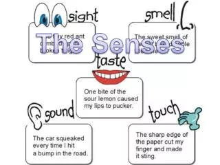
The Senses • General senses of touch • Temperature • Pressure • Pain • Special senses • Smell • Taste • Sight • Hearing • Equilibrium
The Eye and Vision • 70 percent of all sensory receptors are in the eyes • Each eye has over a million nerve fibers • The eye is a sphere about 1 inch in diameter…like a ping pong • Only 1/6 of the eye is seen • Most of the eye is surrounded by bone & cushioned by fat
Accessory Structures of the Eye • Eyelids • Eyelashes • Muscles Figure 8.1b
Accessory Structures of the Eye • Conjunctiva • Membrane that lines the eyelids • Connects to the surface of the eye & secretes mucus for lubrication
Homeo Imbalance • Conjunctivitis = reddened irritated eyes. “Pinkeye” is the HIGHLY infectious form caused by bacteria or virus
Accessory Structures of the Eye • Lacrimal apparatus • Lacrimal gland – produces diluted salt solution (tears) • Lacrimal canals – drains tears from eyes • Lacrimal sac – provides passage of tears towards nasal cavity Figure 8.1a
Properties of lacrimal fluid (AKA TEARS!) • Dilute salt solution which contains lysozyme an anti-bacterial protein • Protects, moistens, and lubricates the eye • Empties into the nasal cavity
Nasolacrimal duct – empties tears into the nasal cavity (connects eye with nose) • Crying makes you sniffle
Homeo Imbalance • A cold or allergies can cause the lacrimal duct to swell shut. This stops drainage of tears and you get watery eyes.
Structure of the Eye • The wall is composed of three tunics (layers) • Fibrous tunic – outside layer • Choroid – middle layer • Sensory tunic – inside layer Figure 8.3a
The Fibrous Tunic • Sclera • White connective tissue layer • Cornea • Transparent allows light to pass through • Vulnerable to damage but repairs itself easily • The only human tissue that can be transplanted without fear of rejection (no blood=no antibodies)
Choroid Layer • Blood-rich nutritive tunic • Pigment dark in color prevents light from scattering • Modified interiorly into two structures • Cilliary body – smooth muscle, focuses lens for clear vision • Iris- smooth muscle, regulates amount of light that enters • Pigmented layer that gives eye color • Pupil – rounded opening in the iris
Sensory Tunic (Retina) • Contains millions of receptor cells called photoreceptors • Rods & Cones • Signals pass from photoreceptors to retina • Signals leave the retina toward the brain through the optic nerve
Homeo Imbalance • Retinal Detachment: Retina separates from choroid. Retina cannot get nutrients and can die. Easily fixed with laser surgery. • Caused by violent motion of the head, genetics
Lens • Biconvex crystal-like structure • Held in place by a ligament attached to the ciliary body Figure 8.3a
The lens divides the eye into 2 segments or chambers • Homeo Imbalance • Cataracts: Occur as we age. The lens becomes hard and opaque
Internal Eye Chamber Fluids • Aqueous humor • Watery fluid found in chamber between the lens and cornea • Maintains intraocular pressure • Provides nutrients for the lens and cornea
Homeo Imbalance • If the aqueous humor cannot drain, pressure in the eye increases dramatically. • This leads to glaucoma, which will become painful and possibly lead to loss of sight. • Early detection is key since a lot of damage can be done w/o pain. • The machine that blows on your eye!
Internal Eye Chamber Fluids • Vitreous humor • Gel-like substance behind the lens that fills the eyeball • Lasts a lifetime and is not replaced (can be floaters)
Neurons of the Retina and Vision • Rods • Most are found towards the edges of the retina • Allow dim light & peripheral vision • all in gray tones • Cones • Allow for detailed color vision • Densest in the center of the retina
Cone Sensitivity • There are 3 types of cones each sensitive to different wavelengths • Total Color blindness is the result of lack all cone types. Partial is due to lack of 1 or 2 types. Figure 8.6
Lens Accommodation • Light is bent or refracted as it enters the eye • The lens changes shape so that the light is focused on the retina • The ability to focus on objects closer is called accommodation • As objects come closer the lens bulges
Vision Problems • Perfect vision is called emmetropia or “harmonious vision” • Nearsightedness- you can see up close but not far away. Picture focuses in front of retina • Farsightedness- you can see far away but not close up. Picture focuses behind retina.
Images Formed on the Retina • The image on the retina is reversed, upside-down and smaller Figure 8.10
Visual Pathway • Optic nerve: carry impulses from retina to brain, • Part of each optic nerve crosses at the optic chiasma • Each side of brain receives info from both eyes • Allows for binocular vision & depth perception Figure 8.11
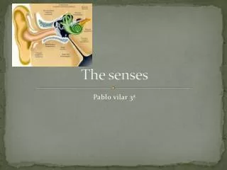
The Ear • Houses two senses • Hearing • Equilibrium (balance)
Anatomy of the Ear • The ear is divided into three areas • Outer (external) ear (Hearing) • Middle ear (Hearing) • Inner ear (Hearing & Balance) Figure 8.12
The External Ear Structures of the external ear • Pinna (auricle)- collects and directs sound waves into the auditory canal • External auditory canal-short chamber in the temporal bone. The walls are covered in ceruminous glands which produce earwax (cerumen). • Tympanic membrane- end of external ear. Vibrates when sound hit it. Figure 8.12
The Middle Ear or Tympanic Cavity • Air-filled cavity within the temporal bone • Two tubes are associated with the inner ear • The opening from the auditory canal is covered by the tympanic membrane • The auditory tube connecting the middle ear with the throat • Allows for equalizing pressure (popping of ear) • This tube is otherwise collapsed
Homeo Imbalance • Otitis Media-Inflammation of the middle ear is a common result of a sore throat. In acute forms the eardrum bulges and becomes inflamed. Fluid needs to be drained with a semi-permanent ear tube and antibiotics. If untreated it can lead to hearing loss.
Bones of the Tympanic Cavity • The 3 bones in the cavity collectively called the Ossicles. • Smallest bones in the body • Malleus (hammer) • Incus (anvil) • Stapes (stirrup) • Transfer and amplify the vibration from the ear drum to the fluid in the inner ear! Figure 8.12
Inner Ear or Bony Labyrinth • Maze of bony chambers in the temporal bone behind the eye socket. • The chambers are filled with perilymph fluid (plasma-like) • Suspended in the perilymph are fluid filled sacs called endolymph Figure 8.12
Inner Ear or Bony Labrynth • 3 subdivisions • Cochlea • Vestibule • Semicircular canals Figure 8.12
Organs of Hearing • Organ of Corti • Located within the cochlea • Contain receptors = hair cells • Above and below cochlear duct contain perilymph. Sound waves set this fluid into motion.
Waves hit the basilar membrane & the hairs on it are bent by the movement of the gel-like tectorial membrane above them. Cochlear nerve attached to hair cells transmits nerve impulses to auditory cortex on temporal lobe
Mechanisms of Equilibrium • Our brains compensate for disturbances in balance • This is a reflex that depends on sensory receptors with in the vestibule and semi-circular canals. • Receptors called vestibular apparatus is divided into 2 functional parts: Static and dynamic equilibrium
Static Equilibrium- “at rest” • Maculae – receptors in the vestibule • Report on the position of the head in respect to gravity • Anatomy of the maculae • Each maculae is a patch of hair cells embedded in the otolithic membrane (gel-like) • Otoliths (tiny stones) float in the gel around the hair cells • Movements cause otoliths to roll and pull the gel which bends the hair cells (creates signal) • The information is sent via the vestibular nerve and then to the cerebellum
Function of Maculae Figure 8.13a–b
Dynamic Equilibrium • Occur in the Semicircular canals. Orientated in 3 planes so no matter the movement, it is detectable. • Responds to angular movements. When dancing or rocking on a boat it goes into over drive. • Within each each semi-circular there is a region called: • Crista ampullaris: Tuft of hair cells covered by the Cupula (gelatinous cap) • Drag and motion of the endolymph is transmitted to the brain Figure 8.14c
neat • When you hear the same tone… • Your auditory receptors tune it out (hum of air conditioning, car motor, etc) • BUT hearing is the last sense to leave our awareness when we fall asleep or receive anesthesia (or even when we die) and the first to come back as we awaken.
Chemical Senses – Taste and Smell • Both senses use chemoreceptors • Stimulated by chemicals in solution • Taste has four types of receptors • Smell can differentiate a large range of chemicals • Both senses complement each other and respond to many of the same stimuli
Olfaction – The Sense of Smell • Olfactory receptors are in the roof of each nasal cavity • Neurons with long cilia (hairs) covered in mucus • Chemicals dissolve into mucus & are detected • Impulses are transmitted via the olfactory nerve and interpretation of smell is made in the brain • Smells create “smell snapshots” which are often linked with emotions. • Reaction to smell is rarely neutral
Olfactory Epithelium Figure 8.17
The Sense of Taste • Taste buds house taste receptor organs • Most are on the tongue (~10K) • Few on soft palate & cheeks
The Tongue and Taste • The tongue is covered with projections called papillae. Give tongue it’s texture • Fungifiorm papillae – rounded projection with taste buds, most numerous • Circumvallate papillae – large papillae with taste buds, few in back • Taste buds are found on the sides of papillae
Structure of Taste Buds • Gustatory cells are the receptors that respond to chemicals in saliva • Have gustatory hairs (long microvilli) • The long microvilli protrude through the taste pore and when stimulated, depolarize and the impulses go to the brain and you taste
Anatomy of Taste Buds Figure 8.18