Download
1 / 73
731 likes | 821 Vues
Explore the bony anatomy of the shoulder and arm with orthopedic surgeon and faculty of medicine at IUG-Gaza, Dr. Fadel Naim. Learn about the regions of the upper limb, functions of the clavicle, cleidocranial dysostosis, and clavicle fractures and complications. Discover ligament and muscle attachments to the clavicle, treatment options, and thoracic outlet syndrome.

E N D
Anatomy of Shoulder and Arm Bony Anatomy Dr. Fadel Naim Orthopedic Surgeon Faculty of Medicine IUG-Gaza
77777777777777777777777777777777777777777777777777777777777777777777777777777777777777777777777777777777777777777777777777777777777777777777777777777777777777777777777777777777777777777777777777777777777777777777777777777777777777777777777777777777777777777777777777777777777777777777777777777777777777777777777777777777777777 لَا أُقْسِمُ بِيَوْمِ الْقِيَامَةِ{1} وَلَا أُقْسِمُ بِالنَّفْسِ اللَّوَّامَةِ{2} أَيَحْسَبُ الْإِنسَانُ أَلَّن نَجْمَعَ عِظَامَهُ{3} بَلَى قَادِرِينَ عَلَى أَن نُّسَوِّيَ بَنَانَهُ{4}
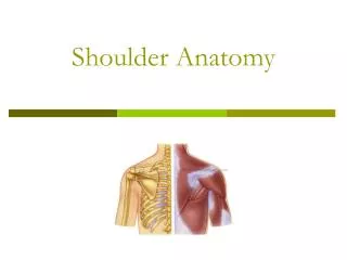
Pectoral Girdle clavicle scapula humerus
Clavicle • The clavicle is an s-shaped bone that attaches the trunk to the upper extremity • Medial 2/3 convex forward and lateral 1/3 concave forward
Clinical Connection – Fractured Clavicle • A fall on an outstretched arm (F.O.O.S.H.) injury can lead to a fractured clavicle • The clavicle is weakest at the junction of the two curves • Forces are generated through the upper limb to the trunk during a fall • Therefore, most breaks occur approximately in the middle of the clavicle
Acromial end • is flat and has a small facet for articulation with the acromion • Sternal end • has a large facet for articulation with the manubrium, and first costal cartilage • Conoid tubercle • Attachment of conoid ligament of the coracoclavicular ligament • Trapezoid line • Attachment of trapezoid portion of the coracoclavicular ligament
FunctionsOfTheClavicle • Serves as a strut from which the scapula and free limb are suspended, keeping them away from the thorax so that the arm has maximum freedom of motion • Forms one of the bony boundaries of the cervico-axillary canal, affording protection to the neurovascular bundle supplying the upper limb • Transmits shocks (traumatic impacts) from the upper limb to the axial skeleton • Provides attachment for muscles Cleidocranial Dysostosis
Ossification Of The Clavicle • The clavicle is the first long bone to ossify, beginning during the 5th and 6th embryonic weeks • Is completed it between the 25th and 31st years. • This is the last of the epiphyses of long bones to fuse. • A smaller scale-like epiphysis may be present at the acromial end of the clavicle; it must not be mistaken for a fracture.
Clavicle: Ligament Attachments • Sternal end of clavicle to first costal cartilage: Costoclavicular ligament • Conoid tubercle: Conoid portion of coracoclavicular ligament • Trapezoid line: Trapezoid portion of coracoclavicular ligament
Clavicle: Muscle Attachments • Deltoid • Pectoralis major • Trapezius • Sternocleidomastoid • Subclavius
Muscular, ligamentous, and fascial attachments to the clavicle
ClavicleFracture • The clavicle is one of the most commonly fractured bones in the body with indirect trauma being the usual cause (Sport injuries) • Midclavicular fractures account for 80% of clavicular fractures, with distal fractures at 15% and proximal fractures at 5%.
ComplicationsofClavicleFracture • Rare but serious neurovascular complication, such as a tear of the subclavian artery or brachial plexus injury, must be kept in mind when evaluating and treating clavicular fractures • Neurovascular examination on initial evaluation is very important • carefully evaluation of: • Pulses in the distal part of the upper extremity • Strength • Sensation
The close relationship of the supraclavicular nerves to the clavicle may result in their involvement in callus formation after fracture of the bone. This may be the cause of persistent pain over the side of the neck.
A fracture through the clavicle causes the shoulder to sag forward and downward. • Because of the weight of the upper limb the trapezius muscle is unable to hold the lateral fragment up so the shoulder drops • The sternodeidomastoid muscle elevates the medial fragment of bone • The lateral fragment of the clavicle may be pulled medially by the adductor muscles of the arm, such as the pectoralis major. • Overriding of the bone fragments shortens the clavicle.
TreatmentandPrognosis • Most fractures of the clavicle heal well • Mid and proximal clavicular fractures are usually treated using figure-of-eight strapping • Immobilization is usually discontinued at 3-4 weeks
The Thoracic Outlet Syndrome • The thoracic outlet contains: • The first rib • The subclavian artery and vein • The brachial plexus • The clavicle • The lung apex. • Injury to these structures may result in postural or movement-induced pain around the shoulder and supraclavicular region • Most of the symptoms are caused by pressure on the lower trunk of the plexus producing pain down the medial side of the forearm and hand and wasting of the small muscles of the hand. • Pressure on the blood vessels may compromise the circulation of the upper limb.
Scapula • Flat triangular bone • On the posterior thoracic wall • Between 2nd and 7th rib
AnteriorScapula • Borders: • Superior • Medial • lateral • Angles: • Superior • inferior • coracoid process • acromion • neck of scapula
Anterior Scapula acromion process coracoid process glenoid cavity superior angle subscapular fossa inferior angle
Subscapular fossa • Origin of the subscapularis muscle • Suprascapular notch • The superior transverse scapular ligament traverses this notch. • The suprascapular artery passes over it • The suprascapular nerve passes under the ligament.
PosteriorScapula • Spine of scapula • Divides the supraspinous and infraspinous fossae • Serves as attachment for the deltoid and trapezius • Acromion: • Lateral extension of spine of scapula; • Articulate with clavicle • Greater scapular notch • Point at which the spine of the scapula ends, but the acromion continues; • Coracoid process • Partially seen as it projects anteriorly;
Supraspinousfossa • Origin of the supraspinatus muscle • Infraspinousfossa • Origin of the infraspinatous muscle • Lateral border • Attachment of: • Teres major • The long head of the triceps brachii • Teres minor
Posterior Scapula acromion process supraspinous fossa infraspinous fossa spine lateral border medial border
LateralScapula • Supraglenoid tubercle • Attachement of the long head of the biceps brachii • Infraglenoid tubercle • Attachement of the long head of the triceps brachii
LateralScapula • Acromion: • Articulates with the clavicle • Attachment for the trapezius and deltoid muscles; • Superior and inferior angles • Coracoid process: • Attachment point for: • The short head of the biceps brachii • Corachobrachialis • Pectoralis minor
Muscular and ligamentous attachments, costal aspect of scapula
Muscular and ligamentous attachments, dorsal aspect of scapula
Fracture Of The Scapula • Many fractures of the scapula result from high-impact accidents • such as being thrown from a motorcycle • 9/10 of those with fractures of the scapula also suffer from life-threatening injuries • rib fractures • lung injuries • head and spinal cord injuries.
Fracture Of The Scapula • The diagnosis of a fracture of the body of the scapula is often overlooked • If the fracture is not associated with other life-threatening injuries • a sling or shoulder immobilizer may be used for 1-to-2 weeks until the pain subsides.
WingedScapula • The serratus anterior muscle(innervated by long thoracic nerve) pulls the medial border of the scapula to the posterior thoracic wall and stabilizes it
WingedScapula • When the serratus anterior is paralyzed the medial border of the scapula moves laterally and posteriorly away from the thoracic wall • This giving the scapula the appearance of a wing, consequently the term “winged scapula”]
Weakness or paralysis of serratus anterior may be secondary to: • Lesions of the 5/6/7 cervical nerve roots (injury or viral neuropathy) • Injury to the brachial plexus • Direct damage to the long thoracic nerve • Weapons, including missiles (bullets) directed toward the thorax, are a common source of injury
A way to check if the muscle is working properly is to have a person push against a wall or door • When the arm is raised, the medial border and inferior angle of the scapula pull markedly away from the posterior thoracic wall • Disability is rare however if function is noticeably impaired • Transfer of sternal portion of pectoralis major via a fascia lata graft to the lower pole of the scapula • The scapula can be fixed to the rib cage
The Humerus • The humerus is the bone of the shoulder and arm. • It articulates with the scapula at the shoulder and with the radius and ulna at the elbow. • The proximal end consists of: • The head • Anatomical neck • Greater and lesser tubercles separated from each other by an intertubercular groove (bicipital groove)
The head • The head, nearly hemispherical in form • Directed upward, medialward, and a little backward • Articulates with the glenoid cavity of the scapula. • The circumference of its articular surface is slightly constricted and is termed the anatomical neck
The anatomical neck • The anatomical neck of the humerus is an indentation distal to the head of the humerus on which the articular capsule attaches. • It is best marked in the lower half of its circumference • In the upper half it is represented by a narrow groove separating the head from the tubercles. • Fracture of the anatomical neck rarely occurs.
The Greater Tubercle • The greater tubercle is situated lateral to the head and lesser tubercle, and just lateral to the anatomical neck • It is covered by the deltoid muscle, which is responsible for the normal, rounded contour of the shoulder. • Its upper surface is rounded and marked by three flat impressions: • the highest for insertion of the suprasinatus muscle • the middle for the infraspinatus muscle • the lowest one, and the body of the bone for teres muscle • The lateral surface of the greater tubercle is convex, rough, and continuous with the lateral surface of the body