Understanding the Reproductive System for Healthy Living
Learn about male and female reproductive systems, embryonic development, and family planning. Explore the anatomy, functions, and hormonal control of reproduction. Prepare for an exam with comprehensive study materials.

Understanding the Reproductive System for Healthy Living
E N D
Presentation Transcript
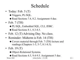
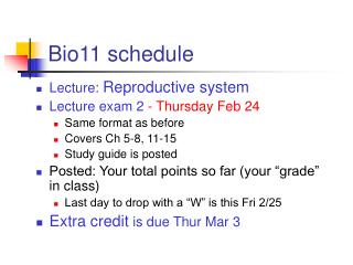
Bio11 schedule Lecture: Reproductive system Lecture exam 2 - Thursday Feb 24 Same format as before Covers Ch 5-8, 11-15 Study guide is posted Posted: Your total points so far (your “grade” in class) Last day to drop with a “W” is this Fri 2/25 Extra credit is due Thur Mar 3
Ch 16 Reproductive System Males & Females Embryonic Development Family planning HIV/AIDS
The male reproductive system • Designed for the continuous production of a large number of sperm • Produces testosterone, the male sex hormone
Sperm cells Highly specialized for their role in fertilization Head: contains the nucleus Acrosome: contains enzymes to digest a passage to the egg Mitochondria: function? Tail: for movement
Formation of sperm • The testes • produce sperm • the hormone testosterone • Sperm do not complete their development at body temp (37°C) • So the testes are outside of the body in the scrotum
Spermatogenesis • The testis is packed with tightly coiled seminiferous tubules • Sperm production, or spermatogenesis, takes place inside the tubules • Spermatogenesis begins in germ line cells on the outside of the tubule
Spermatogenesis • Germ line cells in the seminiferous tubules undergo meiosis to form sperm • Are sperm haploid or diploid? • They are released into the seminiferous tubule • Adult males produce several hundred million sperm each day
Meiosis: review Germ line cell is diploid DNA is replicated Two cell divisions Meiosis I and Meiosis II. The result? 4 haploid daughter cells 2n Produces gametes n
Spermatogenesis • Sperm cells are made in the testis • Develop motility in the epididymis • Delivered to the vas deferens • When sperm is ejaculated, it travels from the vas deferens to the urethra
Spermatogenesis • Sperm leave the penis in a fluid called semen • Various glands (seminal vesicle, prostate gland and Cowper’s gland) add fluids which help nourish the sperm
Sperm delivery system • The penis contains 3 long cylinders of spongy tissue • It is designed to inflate • Nerve impulses cause the blood vessels leading into this tissue to expand • Blood collects in the spongy tissue and causes the penis to become erect and rigid • Continued stimulation is required for ejaculation
Hormones control the male reproductive system • The pituitary gland secretes 2 hormones, FSH and LH • FSH stimulates sperm formation • LH stimulates the testis to produce testosterone FSH LH
Testosterone • Develops male sex characteristics • Enlarges the sex organs • Body hair, beard • Voice change • Develops sexual function • Sex drive (libido) • Sperm maturation • Stimulates bone and muscle growth
The female reproductive system • Designed to • Produce 1 egg each month • Prepare the uterus for implantation of the fertilized egg Ovulation
Anatomy of the female reproductive system • The eggs, or ova, mature in the ovaries • The fallopian tubestransport egg to the uterus • The uterus is lined with epithelial tissue called the endometrium • the surface of the endometrium is shed during menstruation • the uterus narrows to a muscular “neck” called the cervix • The vagina leads from the uterus to the external genitalia
The Female Reproductive Cycle • The female reproductive cycle is actually two cycles in one: • The ovarian cycle • Growth and release of 1 egg each month • Coordinated by FSH and LH • The menstrual cycle • prepares the uterus for possible implantation of an embryo. • Coordinated by estrogens and progesterone
The ovarian cycle • Eggs develop from cells called oocytes • During each reproductive cycle, one (usually) of the oocytes matures • Only ~400 oocytes mature and are ovulated in a woman’s lifetime Ovary and Fallopian tube
Ovulation –only 1 egg matures each month • Ovulation: the mature follicle discharges the egg • The egg is swept up into the Fallopian tube • where fertilization may occur Ovary and Fallopian tube
FSH and LH coordinate the ovarian cycle FSH • FSH stimulates the growth and maturation of a follicle • The follicle cells secrete estrogen into the bloodstream
FSH and LH coordinate the ovarian cycle FSH FSH andLH • Estrogen levels peak at 12 days • this causes LH levels to surge • and stimulates ovulation at 14 days • The mature follicle bursts and releases an egg
FSH and LH coordinate the ovarian cycle FSH FSH andLH LH • LH (luteinizing hormone)stimulates • Formation of the corpus luteum • Secretion of progesterone and estrogens • If the egg is not fertilized, the corpus luteum breaks down
The uterine (menstrual) cycle • Rising levels of progesterone and estrogens promote thickening of the endometrium • When the corpus luteum breaks down drop in levels of these hormones • Endometrium begins to shed – menstruation
28_24a Pituitary FSH LH Ovary Uterus Time
FSH, LH Female reproductive cycle X Negative feedback Estrogen Progesterone Menstrual cycle Ovarian cycle
Estrogens have several important functions • Develop and maintain female reproductive structures, secondary sex characteristics, and the breasts • Increase protein synthesis • Preserve bone density • Regulate synthesis of cholesterol by liver
It all starts with fertilization • Copulation releases hundreds of millions of sperm into the vagina • Only a few hundred survive the trip to the egg • Only one will fertilize it.
The process of fertilization Onlyone sperm reaches the egg Figure 26.13
The egg’s journey • Fertilization takes place in the Fallopian tube • The fertilized egg begins mitosis – a series of cell divisions called cleavage • It eventually forms a ball of cells called a blastocyst which implants in the lining of the uterus
The blastocyst • Implantation occurs at day 7 • The inner cell mass forms the developing embryo • These are pluripotentstem cells – they give rise to all types of cells in the body • The outer cell layer (trophoblast) becomes part of the placenta • Provides nutrients and oxygen to the embryo
What prevents menstruation? • The implanted embryo secretes a hormone, human chorionic gonadotropin (hCG) • hCG tells the corpus luteum to continue secreting estrogen and progesterone • This prevents menstruation • Because hCG comes from the embryo and not from the mother, pregnancy tests measure hCG
Stages of early development • Fertilization • Cleavage • Implantation of the blastocyst (day 7) • Neural tube forms (days 16-25) • the first tissue to develop • Organogenesis (4 weeks) • Formation of body organs • From embryo to fetus 1 4:18 http://www.youtube.com/watch?v=0xvJ-NNuR6M
Fetal Development: organogenesis • The process of forming body organs begins in the 4th week of pregnancy • This is a crucial time • Most spontaneous abortions (or miscarriages) occur during this period • Alcohol use during pregnancy is one of the leading causes of birth defects, producing fetal alcohol syndrome Human embryo at 4 weeks
A month-old embryo • Brain and spinal cord have begun to take shape • Four stumpy limb buds • A short tail • Looks like most month-old vertebrate embryos
Programmed cell death –a key developmental process • Certain tissues produced during embryonic development are destroyed– apoptosis • Cells in the developing hands and feet are killed, separating the fingers and toes
Fetal Development: the first trimester • Development is essentially complete at the end of the third month of pregnancy • All the major organs are present • Arms and legs begin to move • The developing human is now referred to as a fetus Human fetus at 3 months 2 From embryo to fetus 3:20 http://www.youtube.com/watch?v=aw5v6_5GaLQ&NR=1
Ultrasound imaging • By the end of the 1st trimester, the sex of the fetus can be determined by an ultrasound exam • An ultrasound image is produced when high-frequency sound waves are bounced off the fetus
Fetal Development • The second trimester is a time of growth • During the 4th month, the bones enlarge • Mother can feel baby kicking • By the end of month 6, the fetus can survive outside the uterus with special medical care Human fetus at 4 months
Fetal Development • The third trimester is a period of rapid growth • The weight of the fetus doubles • Brain and lungs complete development • The growing fetus is fed by the placenta which passes nutrients from the mother’s bloodstream into the fetal blood supply • But maternal and fetal blood don’t mix
The placenta • An organ unique to mammals • Allows close contact between the bloodstreams of the developing fetus and mother • The placenta supplies the fetus with oxygen and nutrients, and allows fetal waste to be disposed of via the mother’s kidneys • Placenta also synthesizes hormones required to maintain pregnancy Oxygen, nutrients and hormones to baby Wastes and CO2 from baby
Childbirth • The birth of a child is brought about by a series of strong, rhythmic contractions of the uterus called labor • Labor is induced by a strong surge of 3 hormones
Three stages of labor • Dilation of the cervix • Opens to a width of 10 cm (4 inches) • Delivery of the infant • Strong uterine contractions • Delivery of the placenta • The “afterbirth”
Family planning Preventing pregnancy
Birth control • Contraception methods differ in their effectiveness • Most effective • Sterilization (vasectomy, tubal ligation) • IUD • Implant • Least effective • Withdrawal • Spermicides • See Birth control effectiveness chart on Planned Parenthood website • http://www.plannedparenthood.org/health-topics/birth-control//birth-control-effectiveness-chart-22710.htm
Sterilization tubal ligation vasectomy
What is an IUD? • Intrauterine device • IUDs are small, "T-shaped" devices inserted into a woman's uterus to prevent pregnancy. • Effective for at least 5 years • How Does an IUD Work? • Copper affects the motility of sperm, preventing fertilization. • Also alters the lining of the uterus and prevents the fertilized egg from implanting in the uterus.
The implant • A matchstick-sized rod that is inserted in the upper arm • Releases the hormone progestin and prevents ovulation • It protects against pregnancy for up to 3 years.
Other methods that use hormones to prevent ovulation • Hormone methods • Birth control Shot, Pill, Ring, Patch • Breast feeding • The birth control shot contains progestin • The pill, ring and patch contain both estrogen and progestin. • The hormone methods work by preventing ovulation
FSH LH Negative feedback How do these hormones prevent ovulation? • Female sex hormones coordinate the reproductive cycle • FSH and LH Ovulation • Estrogen and progesterone prepare the uterine lining for implantation • Birth control pills contain estrogen and progestin • Turn off production of FSH and LH • Prevent ovulation Estrogen Progesterone
Breastfeeding • While a woman is continuously breastfeeding, her body does not make hormones that are necessary for ovulation • Less than 1 out of 100 women who practice continuous breastfeeding perfectly will become pregnant. • Using breastfeeding as birth control can be effective for 6 months