Download

1 / 30
340 likes | 610 Vues
Flow Cytometric Abnormalities in Myelodysplastic Syndrome. Raida Oudat,MD Consultant Hematopathologist at Princess Iman Research and Laboratory Sciences Center. KHMC. Amman-Jordan. INTODUCTION.

E N D
Flow Cytometric Abnormalities in Myelodysplastic Syndrome Raida Oudat,MD Consultant Hematopathologist at Princess Iman Research and Laboratory Sciences Center. KHMC. Amman-Jordan.
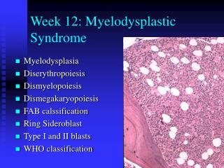
INTODUCTION • The myelodysplastic syndromes (MDS) are clonal stem cell diseases characterized by ineffective hematopoiesis with peripheral cytopenia(s),morphological dysplasia, and increased risk of development of acute myeloid leukemia • The current diagnostic approach to patients with MDS includes: CBC,BM,iron stain and cytogenetics.
The previous use of FC in MDS has been primarily restricted to the characterization of blast cells in secondary acute leukemia following MDS • Myeloid and monocytic dyspoiesis identified as phenotypic abnormalities by FC in MDS correlates with the International Prognostic Scoring System (IPSS), the WHO-adjusted prognostic scoring system (WPSS), transfusion dependency, and time-to progression to advanced MDS/AML, as well as with outcome.
Immunophenotypic aberrancies of myeloblasts,e.g. over- or decreased expression of common myeloid antigens or expression of lineage infidelity markers, such as CD7 and TDT may have independent prognostic impact even if the percentage of blasts in the bone marrow is lower than 5%.
Lately FC has been utilized in diagnosing low grade MDSs which do not display morphologic dyspoiesis or chromosomal aberrations. • FC is also used in Disease monitoring in MDS by sequential analysis of characteristic aberrancies.
patients can also be monitored by FC to determine disease progression (increased abnormalities) or response to therapeutic interventions. Stable FC aberrancies during treatment may spare patients long-term treatment with ineffective drugs with potential toxicity.
NORMAL HEMATOPOIESIS Normal hematopoiesis begins with bone marrow stem cell proliferation giving rise to cells capable of differentiation along multiple lineages. The differentiation of each lineage occurs in a relatively linear fashion through a sequential series of stages, which result in fully functional mature forms.
In disease states alterations in antigenic expression occur, which reflect abnormalities in maturation or function. • These alterations can be used in the diagnosis and monitoring of the disease.
Blasts in MDS can show increased fluorescence intensity for CD117, CD13 or CD33, decreased CD45 and CD38, or aberrant expression of lymphoid antigens such as CD2, CD5, CD7, and CD56, and paradoxical expression of mature myeloid antigens, such as CD65, CD15, CD10, CD11b.
Hypogranularity in maturing myeloid cells can show decreased side scatter. • Monocytes can show increased numbers, decreased CD14, CD64, CD33, CD11b, CD13, or aberrant CD56 and CD2 expression. • Maturing myeloid cells can also show altered maturation patterns of CD13/CD16 and CD11b/CD16 and, or decreased fluorescence intensity of mature myeloid antigens such as CD10, CD15, and CD33
conclusion • The use of FC in the diagnosis of MDS can be applied with standardized panels and protocols using the most diagnostically useful abnormal antigen patterns. However large prospective clinical studies must be completed to better define the importance of each abnormality that is easy to interpret, reproducible within and between laboratories, and encodes the maximal information for assisting diagnosis.