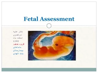
Fetal Assessment
Fetal Assessment. Presented by: Ann Hearn, MSN, RNC 2013. Ultrasound. Definition -- an instrument which uses high frequency sound waves that deflect off of tissue and return as echoes to visualize structures in the body. Ultrasound. Advantages: Results are immediate

Fetal Assessment
E N D
Presentation Transcript
Fetal Assessment Presented by: Ann Hearn, MSN, RNC 2013
Ultrasound • Definition -- an instrument which uses high frequency sound waves that deflect off of tissue and return as echoes to visualize structures in the body
Ultrasound Advantages: • Results are immediate • Requires about 20 - 30 minutes • Allows the mother and family to “see” the baby • NOW IN 3D Disadvantages: • Expensive • No Dx of inborn errors of metabolism
Ultrasound First Trimester (1-12 wks) Transvaginal US –procedure • Empty bladder • Lithotomy position Assessment: • Confirms pregnancy , viability & location • Estimates gestational age • Identify fetal abnormalities • Adjunct to chorionic villus sampling
Ultrasound 2nd & 3rd Trimester (13-40wks) Transabdominal US • Left tilt position with knees sl. Bent • Full bladder (2nd trimester) Assessment: • Confirm viability, estimate gestational age • Evaluate anatomy and placental location • Assess growth • Evaluate amniotic fluid volume • Guide for amniocentesis
Alpha - Fetoprotein (AFP) • Measurement of protein produced by the yolk sac and liver found in fetal plasma. • Elevated AFP may indicate: • Open neural tube defects • Anterior abdominal wall defects • Multiple gestation • Advanced gestational age • Low AFP associated with: • Down syndrome (trisomy 21) • Edwards syndrome (trisomy 18)
Chorionic Villus Sampling(CVS) • Invasive procedure • Removal of small tissue specimen from the fetal portion of the placenta • Tissue obtained about 10 - 13 weeks gestation • Detects chromosomal, metabolic & DNA abnormalities
Chorionic Villus SamplingCVS • Risks: • Failure to obtain tissue • Rupture of amniotic membranes • Leakage of amniotic fluid • Vaginal bleeding • Intraurterine infection • Rh Alloimmunization • Maternal tissue contamination of the specimen • Increased risk of spontaneous abortion
Chorionic Villus SamplingCVS • Nursing interventions • Monitor : • vital signs • FHR • uterine contractions/cramping • vaginal discharge • Administer Rhogam if indicated • Teach patient to report: • Uterine contractions • Vaginal discharge • S/S of infection
Amniocentesis Aspiration of amniotic fluid by insertion of a needle through the abdominal and uterine wall into the amniotic sac
Amniocentesis Purposes: • 2nd trimester • Chromosomal abnormalities • Fetal Rh sensitization • Dxamnionitis • Confirm abnormal AFP (AFAFP) • 3rd trimester • Fetal lung maturity • L/S ratio • Fetal hemolytic disease
Amniocentesis • An invasive procedure • Requires a consent form to be signed • Performed: 2nd trimester (between 15 20 wks gestation ) & during 3rd trimester. • Complications • Trauma • Infection • Hemorrhage • Preterm labor
Amniocentesis • Preparation • Vital Signs and FHT’s • Empty bladder • Abdominal prep and scrub • Ultrasound • Left tilt position • Area of insertion is anesthesized and a needle inserted into the amniotic cavity • 15 - 20 cc of fluid withdrawn for analysis
Amniocentesis • Post care / Discharge Teaching • Monitor V/S, FHT’s and UC’s X 1 hour • Administer Rhogam if Rh negative • Observe for leakage of fluid from site • Teach patient to report – • Fetal hyperactivity or lack of fetal movement • Vaginal discharge: clear or bleeding • Uterine contractions or abdominal pain • Fever or chills
L/S Ratio Lecithin /Sphingomyelin Ratio Lecithin and Sphingomyelin are two components of Surfactant. Assesses Fetal Lung Maturity
L/S Ratio Lecithin /Sphingomyelin Ratio • As surfactant increases in the fetal lungs, the levels of lecithin should also increase. • Lecithin becomes 2 - 3 times > sphingomyelin by about 35 weeks • Fetal maturity & adequate surfactant = L/S ratio > 2 : 1
Karyotyping Determine sex of the fetus Normalcy of Chromosomes
Karyotyping • Indications: • Maternal age 35 or > at time of birth (AMA) • Pervious child born with a chromosomal abnormality • Mother carrying an X-linked disease • Parents carrying an inborn error of metabolism • Both parents carrying an autosomal recessive disease • Family history of neural tube defects
Karyotyping • Trisomy Monosomy
Antepartum Testing • Purpose • Determine fetal health or compromise • Guide interventions • Reduce perinatal morbidity/mortality
Non-Stress Test - NST Assessment of fetal status • Observation of fetal heart rate associated with fetal movement. • The FHR should increase or accelerate with fetal movement • FHR accelerations indicate an intact CNS and adequate oxygenation
Procedure for an NST • Electronic fetal monitor is applied • Fetal movements are documented • Compare the FHR with the fetal movements • Results: • Reactive -- at least two accelerations of FHR with fetal movement of 15 BPM, lasting 15 seconds or more, over 20 minutes. • Nonreactive-- the reactive criteria are not met. Indication of need for further assessment
Contraction Stress Test – CST • A means of identifying the fetus that is at risk for intrauterine asphyxia. Determines utero-placental insufficiency.
Procedure for an CST • Electronic fetal monitor attached • IV Oxytocin (low dose) –or- • Nipple stimulation started • Goal -- 3 contractions of good quality, lasting 40-60 seconds over a 10 minute period
Contraction Stress Test - CST • Results: • Negative -- 3 contractions in 10 minutes with NO signs of late decelerations • Positive -- repetitive persistent late decelerations occurring with more than half the contractions • Equivocal – FHR decelerations with uterine hyperstimulation • Unsatisfactory – fewer than 3 contractions in 10 minutes
Contraction Stress Test • Post CST Monitoring • FHR • Labor • SROM • Discharge instructions • Notify HCP for the following: • Regular painful contractions • Leakage of amniotic fluid • Decrease or increase in fetal movement • Vaginal bleeding
Fetal Assessment Non-Stress Test Reactive Non - Reactive Repeat in 1 - 2 weeks ReactiveStimulate Non- Reactive Contraction Stress Test Negative Positive Further Evaluation Repeat NST in 1 week Possible Delivery
Try This! • Which of the following is NOT an indication of fetal distress? • A reactive non-stress test • Non-reactive non-stress test • A positive CST • A negative CST
Biophysical Profile • Comprehensive assessment of five biophysical variables: • Fetal breathing movement • Fetal movements of body or limbs • Fetal tone (extension and flexion of extremities) • Amniotic fluid volume – visualized as pockets around the fetus • Reactive FHR with activitity (reactive NST)
Biophysical Profile By combining these five assessments, the BPP helps to identify the compromised fetus and to confirm the healthy fetus Since it combines several assessments, it is a better indicator of fetal well-being
Biophysical Profile • A score of 0 or 2 is assigned to each finding for a maximum score of 10. • Scores of 8-10 are considered normal • Lower scores are associated with a compromised fetus and warrant further assessment and possible delivery of the fetus.
Fetal Movement: Kick Counts • Non-invasive • Goal: • 10 kicks in 12 hours • 2-3 times/day – at least 3 movements in 60 minutes
The End Return