Download
1 / 98
1.2k likes | 2.58k Vues
ASSESSMENT OF Fetal Well-being. Lectures 3. Assessment of fetal well-being. The major expected outcome is the detection of potential fetal compromise Used before intrauterine asphyxia of the fetus and health care provider can take measures to prevent or minimize adverse perinatal outcomes

E N D
ASSESSMENT OF Fetal Well-being Lectures 3
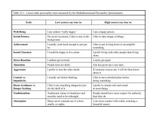
Assessment of fetal well-being • The major expected outcome is the detection of potential fetal compromise • Used before intrauterine asphyxia of the fetus and health care provider can take measures to prevent or minimize adverse perinatal outcomes • First- and second-trimester antepartal assessment is directedprimarily at the diagnosis of fetal anomalies. • Thegoal of third-trimester testing is to determine whether theintrauterine environment continues to be supportive tothe fetus.
Daily Fetal Movement Counts Kick Counts • Assessment of fetal activity by the mother • Non-invasive, inexpensive, simple to understand, and does not interfere with routine at home • once a day, roughly at the same time every day in a comfortable sitting or lying position • when baby is usually active (after meals, after activity, and in the evening). • Since healthy babies have sleep cycles, baby may not kick, or kick less than usual, or have less than 10 kicks in 2 hours. If so, wake up the baby by drinking fluid or by walking for 5 minutes. Repeat the kick count.
Daily Fetal Movement Counts Kick Counts • No less than3 movements in 30 minutes • Most healthy babies should take less than 2 hours for 10 kicks. • not moved 10 times in 2 hours or the baby has sustained significant changes. • Fetal alarm signal if no movement in 12 hours • The evaluation may include: • Ultrasound - taking pictures from sound waves to evaluate the growth of the baby, amniotic fluid quantity, placenta, blood flow pattern etc. • Non stress test (NST) -Baby's heart rate monitoring in response to its own movements • Biophysical profile (BPP) -using an ultrasound exam with a non stress test (NST) to evaluate baby's heart rate, breathing, body movement, muscle tone, and amniotic fluid quantity • Contraction stress test (CST) -Baby's heart rate monitoring in response to uterine contractions
Ultrasound in obstetrics can provide good information about the fetus and its environment With ultrasound, can be determined an early intervention or conservative management in pregnancy Latest developments in ultrasound examination is a transvaginal ultrasound discovery - the observation of "FLOW DOPLLER" and the most sophisticated ultrasound 3 D and 4D which has a high ability to determine fetal condition • 7
Ultrasonography • Indications for use • Fetal heart rate activity • Gestational age • Fetal growth • Fetal anatomy • Fetal genetic disorders and physical anomalies • Placental position and function • visual assistance to other invasive tests • Fetal well-being
Ultrasonography • Abdominal • After 1 trimester • Full blader • Vaginal • 1 trimester • Early diagnostic of uterine pregnancy • Empty blader • Obese woman
Ultrasonography • An important and safe technique in antepartum fetal surveillance • Levels of ultrasonography: • Standard examination • Used for specific indications, i.e., fetal viability, fetal presentation, gestational age, locate the placenta, fetal anatomy and malformation • Specialized or targeted examination • Suspicion of an abnormal fetus (abnormal finding on clinical examination, poly- or oligohydramnionios, elevated AFP)
Ultrasonography: Indication for use 1 trimester • Number, size, location of gestational sac • Fetal cardiac and body movement • Uterine abnormalities (bicornuate uterus, uterine fibroid, IUD) or adnexal masses • Duration of pregnancy (crown-rump length) • Visualization during chorionic villus sampling
Ultrasonography: Indication for use ( 2nd and 3rd trimester ) Fetal viability, number and presentation, Establishment of fetal age and growth by fetal biometry including: BPD ~ biparietal diameter FL ~ femur length AC ~ Abdominal circumference Biophysical profile Evaluation of fetal anatomic structures: Cerebral lateral ventricles Spine Four chamber view of the heart Stomach-bowel,abdominal wall at the area of the umbilical cord insertion Bladder and kidney Limbs and umbilical cord Amount of amniotic fluid Placental localization and maturity Evaluation of the uterine, and adnexae for abnormalities and masses Cervical length Visual assistance to invasive tests • 12
Fetal heart activity • 6-7 weeks by echo scaner • 10-12 weeks by Doopler Fetal viability • Fetal cardiac activity • Fetal movement • Breathing movement
Gestational age • Gestational sac dimensions (about 8 weeks) • Crown-rump length (7-12 weeks) • Biparietal diameter (after 12 weeks) • Femur length (after 12 weeks) • BPD, FL and AC the most important parameters for determination of gestational age • Determination of gestational age should be performed prior to 26 weeks gestational age • 3rd trimester determination of gestational age does not acurately reflect gestational age
Fetal growth • BPD ~ biparietal diameter • FL ~ femur length • AC ~ Abdominal circumference • Discrepancy resulting from inaccurate dates • True intrauterine growth restriction (IUGR) • Symmetric - the fetus being small in all parameters, reflects a chronic or long-standing insult and may be caused by low genetic growth potential, intrauterine infection, undernutrition, heavy smoking, or chromosomal aberration. • Asymmetric - head and body growth varying, suggests an acute or late-occurring deprivation, such as placental insufficiency resulting from hypertension, renal disease, or cardiovascular disease. • Macrosomia - weighing more than 4000 g, associated with maternal glucose intolerance, carries an increased risk of intrauterine fetal death, and at increased risk for trauma during birth.
Ultrasonography: gestational age a In decreasing order b Only if cephalic index ( BPD divided by occipital-frontal diameter ) is normal ( 76-84%) ; otherwise , the fetal head may be dolichocephalic or brachycephalic • 16
1st trimester fetus CRL • 17 8/20/2014
28 mm CRL in 10 weeks twin pregnancy • 18 8/20/2014
Biparietal Diameter a cross section through the fetal head at the level of the thalamus. The skull is represented by the thick white lines which surround the brain. This view is used to measure the biparietal diameter (line) and the circumference of the head (dots). • 19 8/20/2014
Fetal Femur • 20 8/20/2014
Pregnant uterus - longitudinal • 21 8/20/2014
Fetal : intracranial structure and extremity • 22 8/20/2014
Fetalanatomy • Head (ventricles, blood vessels) • Neck • Spine • Heart • Stomach • Small bowel • Liver • Kidney • Blader • Limb
Fetal Cardiac Structure • 24 8/20/2014
Fetal Liver and Lung interface • 25 8/20/2014
Fetal Liver 3rd Trimester • 26 8/20/2014
Fetal Spine • 27 8/20/2014
Spine3 D • 28 8/20/2014
Neural tube defects NTD’s result from failure of tube closure by the 6th weeks gestational age (embryonic age 26 – 28 days ) Various NTD’s anomalies : Anencephaly Encephalocele Spina Bifida • 29
Gross malformation may be detected in 1st trimester sonogram 1: Anencephalus (absence of a major portion of the brain, skull, and scalp) Acrania (partial or complete absence of the cranium). • 30
31 8/20/2014
Spina Bifida Consist of a hiatus, usually in the lumbosacral vertebrae, through which a meningeal sac may protruded → meningocele 90% of cases, the sac contains neural elements→ meningomyelocele The fetal spine should be examined by sonography with: sagittal, tranverse and coronal views • 32 8/20/2014
Spina Bifida • 33 8/20/2014
NEURAL TUBE DEFECTS • 34 8/20/2014
NEURAL TUBE DEFECTS • 35 8/20/2014
Fetal anatomyGross malformation may be detected in 1st trimester sonogram 2: Hydrancephaly (the cerebral hemispheres are absent and replaced by sacs filled with cerebrospinal fluid Cystic Hygroma (is a congenital multiloculated lymphatic lesion that can arise anywhere, but is classically found in the left posterior triangle of the neck. • 36
Abdominal Wall Defects The two most common are : Omphalocele Gastroschisis Can be ascertained early in pregnancy by maternal serum alphafetoprotein screening programs • 37 8/20/2014
Gross malformation may be detected in 1st trimester sonogram 3: Omphalocele (abdominal wall defect in which the intestines, liver, and occasionally other organs remain outside of the abdomen in a sac) Gastroschisis (paraomphalocele congenitalabdominal wall defect in which the intestines and sometimes other organs develop outside the fetal abdomen through an opening in the abdominal wall) • 38
GASTROSCHISIS • 40 8/20/2014
Duodenal atresia Diagnosed prenatally by the demonstration of the double bubble sign( distension of the stomach and first part of the duodenum ) Must be differentiated from other cystic structures in the upper abdomen Diagnosis generally is not possible before 24 weeks 30% of cases has been associated with trisomy 21 • 41 8/20/2014
Gross malformation may be detected in 1st trimester sonogram 4: Fused twins (Siamese twins) are identical twins whose bodies are joined in utero • 42
Fetal genetic Disorders Nuchal Translucency The maximum thickness of the subcutaneus translucent area between the skin and the soft tissues overlying the posterior aspect of the cervical spine in sagital scane plane. (10-14 weeks) A thickness > 3 mm ( sagital plane): 90% trisomy 18 and 13 80% trisomy 21 5% normal • 43
Placenta position and function • Location • Relationship between cervical os • Maturation
Uterus and Adnexa Cervical incompetence : Tunneling of the internal ( dilatation ) Cervical length < 3 cm Bulging membranes ( with or without prolaps of the cord or fetal parts ) 30 weeks of gestational age: length of cervix more than 3 cm Adnexal mass : Physiological: Diameter corpus luteum at pregnancy about 2 cm Uterine fibroid • 45
Sonographic assesment of the amniotic fluid Normal : at 2nd and 3rd trimester vertical pocket about 2 cm AFI ( amniotic fluid index ): sum of the depth of the largest pocket of fluid in the four quadrants of abdomen AFI < 5 cm : strongly asociated with oligohidramnions postmaturity • 46
Amniotic Fluid Index • 47 8/20/2014
Interpretation of the AFl 10.1 to 24.0 cm Normal 5.1 to 10.0 cm Borderline Less than or equal 5.0 cm Abnormal (Oligohydramnios) Greater than 24.0 cm Abnormal (Polyhydramnios) Oligohydramnios is associated with congenital abnomalies (ex. renal agenesis) growth restriction fetal distress Polyhydramnios is associated with neural tube defects obstruction of the fetal gastrointestinal tract, multiple fetuses and fetal hydrops
DOPPLER VELOCIMETRY The primary use of Doppler echo shifts in obstetrics have been to detect and measured blood flow Basis of Doppler Velocimetry : The sound of moving blood cells within vasculature generates an effective Doppler Shift There are 2 methods of estimating circulatory hemodynamics : Direct measurement of the volume of blood flow Indirect estimation of flow velocity using wave form analysis • 49
DOPPLER VELOCIMETRY • The shifted frequencies can be displayed as a plot of velocity versus time, and the shape of these waveform can be analyzed to give information about blood flow and resistance in a given circulation • Velocity waveforms from umbilical and uterine arteries, reported as systolic/diastolic (S/D) ratio achieve