Download

1 / 20
200 likes | 513 Vues
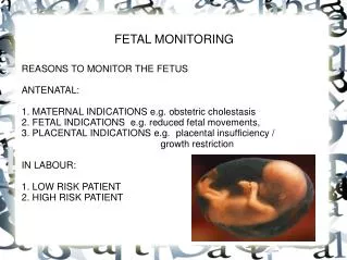
FETAL MONITORING. REASONS TO MONITOR THE FETUS ANTENATAL: 1. MATERNAL INDICATIONS e.g. obstetric cholestasis 2. FETAL INDICATIONS e.g. reduced fetal movements, 3. PLACENTAL INDICATIONS e.g. placental insufficiency / growth restriction IN LABOUR: 1. LOW RISK PATIENT

E N D
FETAL MONITORING REASONS TO MONITOR THE FETUS ANTENATAL: 1. MATERNAL INDICATIONS e.g. obstetric cholestasis 2. FETAL INDICATIONS e.g. reduced fetal movements, 3. PLACENTAL INDICATIONS e.g. placental insufficiency / growth restriction IN LABOUR: 1. LOW RISK PATIENT 2. HIGH RISK PATIENT
ANTENATAL FETAL MONITORING BIOPHYSICAL PROFILE USS: 1. Breathing - Does the baby have breathing movements at least once in 30 minutes? 2. Body Movement - Does the baby move at least three times in 30 minutes? 3. Muscle Tone - Does the baby have at least one flexion-extension (open-close) movement of arms, legs or hands in 30 minutes? 4. Amount of amniotic fluid - Is there enough fluid around the baby? 5. CTG: Is it reactive? AMNIOTIC FLUID The Amniotic Fluid Index (AFI) can be used to determine fetal well-being. Most of the fluid in amniotic fluid is contributed to by fetal urine. This is then resorbed by the membranes and umbilical cord Rapid turnover - possible to measure amniotic fluid from one day to the next
BIOPHYSICAL PROFILE SCORE 8-10 = maximal score 0-4 = severe fetal compromise; delivery indicated
Doppler blood flow velocity waveforms Non-invasive velocity measurements of blood flow Fetus is completely dependent on the supply of oxygen and nutrients from the placenta Examination of the blood flow through the umbilical circulation can assess fetal health Increased placental vascular resistance, reduces velocity of the end-diastolic flow in the umbilical cord artery Several Doppler indices have been used to quantify abnormalities in umbilical artery Doppler flow waveforms: A/B ratio, the resistance index, the pulsatility index Placental insufficiency can be quantified based on the reduction of end-diastolic Doppler flow velocity into (1) reduced enddiastolic flow velocity, (2) absent end-diastolic flow velocity, and (3) reversed end-diastolic flow velocity.
Doppler blood flow velocity waveforms Middle cerebral artery peak-systolic flow velocity (MCA-PSV) use Doppler to detect fetal anaemia
Ductus Venosus Dopplers • May be used as a trigger for delivery of IUGR fetus. • Late sign of CV decompensation • Reflects decreased ability to handle venous return. • Precedes FHR decels • Present in 79/211 (37%) of preterm IUGR, useful > 29wks • Predictive of pH<7.2 • Baschat, O&G, 2007
MONITORING IN LABOUR Intermittent auscultation recommended for low-risk women in established labour INDICATIONS FOR continuous EFM: 1. meconium-stained liquor, 2. abnormal FHR detected by intermittent auscultation 3. maternal pyrexia 4. fresh bleeding in labour 5. oxytocin use for augmentation 6. the woman’s request.
FETAL PHYSIOLOGY 1. The fetal heart pumps deoxygented blood to the placenta via the two umbilical arteries 2. At the placenta there is a free exchange of blood gases (there's no mixing of foetal/maternal blood) 3. The blood is pumped back to the fetus via a single umbilical vein
FETAL HEART RATE The fetal heart is regulated by: 1. Nerve supply i.e. HR is reduced by vagus nerve (parasympathetic), increased by sympathetic supply 2. Circulating catecholamines 3. Central nervous system activity These are influenced by changes in: 1. fetal BP 2. fetal blood gas levels (O2, CO2, pH) 3. Hypoxia 4. Pyrexia 5. Drugs 6. Gestation 7. Cord compression 8. Cerebral activity
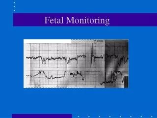
A: Fetal heartbeat; B: Indicator showing movements felt by mother (caused by pressing a button); C: Fetal movement; D: Uterine contractions
Definition of normal, suspicious and pathological FHR traces Classifications of CTG’S 1) Normal: Implies fetal well-being 2) Suspicious: Indicates continued observation /additional tests 3) Pathological: Mandatory Action.
SMALL GROUP / PAIR WORKSHOP using FRESH EYES LABELS
DR. C BRAVADO Define Risk: Low or High Contractions: Frequency, Length Baseline Rate: Bradycardia, Normal, Tachycardia Variability: 5-10bpm/min Accelerations: Present or Absent Decelerations: Present or Absent, Type Outcome: Normal, Suspicious. Pathological. Management Plan
APGAR SCORES DESIGNED TO ASSESS WHICH BABIES NEED RESUSCITATION; IT DOESN'T TELL US WHY A BABY NEEDS RESUSCITATION
CORD GASES Indication of: 1. how well the oxygen supply has been maintained to the fetus during labour 2. How well the fetus has eliminated the waste product CO2 Gives an indication of the efficiency of placental gas exchange during labour Cord gases can suggest a baby has been deprived of oxygen during labour but it cannot tell us if the baby has suffered harm as a result A baby could have good Apgars despite abnormal cord gases A baby that has been deprived of oxygen during labour may have compensated well but is still at risk of of e.g. hypoglycaemia
SMALL GROUP WORKSHOP Divide up into 4 groups Read through the case history Using DR C BRAVADO review the CTG at the times indicated in BOLD