Download

1 / 39
400 likes | 424 Vues
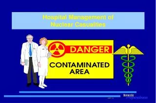
Hospital Management of Nuclear Casualties. Terminal Objective. Recognize the various types of radiological hazards, understand the acute health effects from radiation exposure, hospital procedures for managing exposed and/or contaminated patients, and know how to use radiation protection.

E N D
Terminal Objective Recognize the various types of radiological hazards, understand the acute health effects from radiation exposure, hospital procedures for managing exposed and/or contaminated patients, and know how to use radiation protection.
Terrorist Use of Nuclear Material • Terrorist use of nuclear materials • Simple radiological device • Radiological dispersal device • Reactor • Improvised nuclear device • Nuclear weapon
The Basics of Radiation • Ionizing radiation is electromagnetic energy or energetic particle emitted from a source. • Ionizing radiation is able to strip electrons from atoms causing chemical changes in molecules.
The Basics of Radiation Biological Molecular Damage Biological Damage Chemical Damage Free Radicals 10-10 Seconds Cells, tissues, whole animals Hours to years 1. Proteins 2. Membrane 3. DNA Seconds to hours
Ionizing Radiation - Alpha • 2 neutrons and 2 protons • Highly ionizing • Travels several centimeters in air and a few microns in tissue • Component of nuclear fallout • Stopped by a thin paper or clothing • Threat is inhalation or absorption of alpha emitter in wounds
Ionizing Radiation - Beta • High energy “electron” emitted from nucleus • Can have wide range of energies depending upon the particular radionuclide • Moderately penetrating • Up to a few meters in air • Millimeters in tissue • Some protection by PPE
Gamma or X-Ray (Photons) • High energy rays • Very penetrating • Difficult to shield • Can be produced from radioactive decay and a nuclear weapon explosion or reactor accident • PPE will not protect against photon radiation
Ionizing Radiation - Neutrons • Neutral particle emitted from the nucleus • Can be very penetrating • Requires special consideration for shielding
Radiation Detection • Instrumentation • G.M. Survey Meter • Dose Rate Meter - Ionization Chamber • Alpha Meter • Neutron Meter • Personal Dosimeters • Film Badge • Thermoluminescent Dosimeter • Quarts Fiber Dosimeter • Electronic Instantaneous Read Out Dosimeter
Radiation - Units of Measure • rad - basic unit for measuring radiation • rem - quantifies the amount of damage that is suspected from a particular type of radiation dose
Radiation - Units of Measure X-RAY 100 rads = 100 rem BETA 100 rads = 100 rem GAMMA 100 rads = 100 rem NEUTRONS 10 rads x QF10 = 100 rem ALPHA 5 rads x QF20 = 100 rem
Examples of Radioactive Materials SubstanceHalf LifeEmitUse Americium 241 458 years a, g Smoke Detectors Cobalt 60 5.3 years b, g Medical Therapy Plutonium 238 87 years a Thermoelectric Gen. Plutonium 239 24,400 yrs a Reactors and Weapons Radium 226 1,620 yrs a Medical Therapy Uranium (natural) millions yrs a,b, g Reactors and Weapons Iridium 192 74 days b, g Industrial Radiography
Radiation Half-Life • Time required for a radioactive substance to lose half of its radioactivity • Each radionuclide has a unique half-life • Half-lives range from extremely short (fraction of a second) to millions of years Examples: Tc-99m 6.0 hrs I-131 8.05 days Co-60 5.26 yrs Sr-90 28.1 yrs Pu-239 24,400 yrs U-238 4,150,000,000 yrs
Natural Background Radiation Natural background and manmade radiation 360 mrem Diagnostic chest x-ray 10 mrem Flight from LA to Paris 4.8 mrem Barium enema 800 mrem Smoking 1.5 ppd - 1 year dose 16,000 mrem Heart catheterization 45,000 mrem Mild acute radiation sickness 200,000 mrem LD50 for irradiation 450,000 mrem mrem = millirem = 1/1000 of a rem
Radiation Protection Principles • Time • Distance • Shielding • Quantity
Types of Radiation Injury • External irradiation - whole-body or partial-body • Contamination by radioactive materials - external (deposited on the skin) or internal (inhaled, swallowed, absorbed through skin, or introduced through wounds) • Incorporation of radioactive materials - uptake by body cells, tissues, or organs (bone, liver, kidney, etc) • Combined radiation injury - combination of the above complicated by trauma.
Radiation Injury - Contamination External Internal
Radiation Injury - Incorporation Thyroid Lung Liver Bone
Radiation - LD50 • We know what radiations are produced • We know how to measure them • But the body senses cannot detect radiation. Therefore, how can we measure the biological damage? • LD50/30 Animals • LD50/60 Man
Examples of LD50 for Given Species SpeciesDose (rads) • Guinea Pigs 250 LD 50/30 • Goat 350 LD 50/30 • Man 250-450 (LD 50/60) • Mouse 570 LD 50/30 • Rat 550-800 LD 50/30 • Frog 700 LD 50/30 • Snail 8,000-20,000 LD 50/30
Severity of Injury The higher the dose, the more severe the early effects and the greater the possibility of delayed effects
Survival Time Survival Time Hematopoietic Gastrointestinal CNS/ CVS 200 Rads 2 Gy 1000 Rads 10 Gy 100,000 Rads 100 Gy
Acute Radiation Syndrome (ARS) • Group of symptoms that develop after total body irradiation (> 100 rads) • May occur from either internal or external radiation • Four important factors are: • High Dose • High Dose Rate • Whole Body Exposure • Penetrating Radiation
ARS - Phases • 1. Prodromal Phase - occurs in the first 48 to 72 fours post-exposure and is characterized by nausea, vomiting, and anorexia. At doses below about 500 rads last 2 to 4 days. • 2. Latent Phase - follows the prodromal phase and lasts for approximately 2 to 2 1/2 weeks. During this time, critical cell populations (leukocytes, platelets) are decreasing as a result of bone marrow insult. The time interval decreases as the dose increases. • 3. Illness Phase - period when overt illness develops • 4. Recovery or Death Phase - may take weeks or months
ARS - Hematopoietic Syndrome 3.0 2.5 Normal Range 2.0 Absolute Lymphocytes (109/L) 1.5 Moderate 1.0 Severe Patient Injury 0.5 Very Severe 0.1 Lethal 0 3 6 17 24 48 hrs
ARS - Blood Count RBC Cell Reduction Neutrophils Lymphocytes Platelets 24-hr 1 week 2 weeks 3 weeks
ARS - Gastrointestinal Syndrome • Radiation > 600 rads • Damages intestinal lining • Nausea and vomiting within the first 2 - 4 hours • May develop diarrhea • Associated with sepsis and opportunistic infections • At 10 days could develop bloody diarrhea resulting in death
ARS - Central Nervous System • Seen with radiation dose > 1,000 rads • Microvascular leaks Õ edema • Elevated intracranial pressure • Death within hours
ARS - Skin Dry Desquamation Moist Desquamation Erythema Necrosis Epilation Response 300 600 1000 >1500 >5000 Dose
ARS & Trauma • Radiation and Trauma = á Mortality • Wound and burn care, surgery, and orthopedic repair should be done in the first 48 hours or delayed for 2 to 3 months Emergency Surgery Hemopoietic Recovery No Surgery Surgery Permitted 24 - 48 Hours After 3 Months 3 Months
Triage • Triage: • Use your own triage method • Ensure ABC’s • Stabilize the patient first and only when this is done does one consider irradiation and contamination.
Classification, Treatment & Disposition • Patients are classified in three categories based on signs and symptoms: • Survival probable < 100 rads • Survival possible 200 - 800 rads • Survival improbable > 800 rads
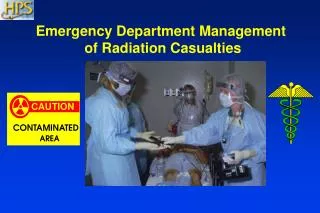
Evaluation & Treatment - Hospital Care • Activate hospital plan • Establish triage area • Plan to control contamination • Prepare area by cover/marking floor, control ventilation • Prepare staff by issuing protective clothing • Prepare for surveying • Establish area for storage of waste • Plan for decontamination of non-traumatized patients
Hospital Care - Patient Arrival • Carefully remove and bag victim’s clothing and personal belongings • Survey patient and conduct biological sampling • Contaminated patients require decontamination • If patient has a wound, decontaminate it first, then decontaminate skin
Decontamination • Irrigate open wounds and cover with sterile dressing • Soap and water showering (including hair) • Effective for mixed radiation/chemical contamination • Refer for any surgery
Internal Contamination/Incorporation • Various medications can be used to limit uptake or facilitate removal of radioactive material • Numerous medications are approved by the FDA. Certain drugs are investigational and can be used in an emergency (i.e. Radiogardase [Prussian Blue] and DTPA) • NCRP 65
Psychological Casualties • Mass Casualty Incident (MCI) caused by nuclear terrorism will create large numbers of concerned casualties who may not actually be injured or contaminated • Establishing counseling centers will prevent psychological casualties from overwhelming health care facilities
Key Points • Pre-planning to ensure adequate supplies of PPE and survey instruments • Training to ensure competence at all levels • Rescue and treatment protocols vary little for radiation contamination • Donning PPE and decontaminating patients minimizes exposure risk • Early symptoms are an indication of the severity of the radiation dose • Treatment requires a unified effort