Anatomy of the Duodenum and Pancreas
Discover the intricate details of the duodenum and pancreas, including parts, relations, mucous membrane features, blood supply, lymph drainage, and pancreatic functions.

Anatomy of the Duodenum and Pancreas
E N D
Presentation Transcript
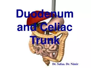
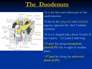
The Duodenum • It is the first and widest part of the small intestine. • It lies in the epigastric and umbilical regions, opposite the first 3 lumbar vertebrae. • It is a C-shaped tube, about 10 inch. It has 4 parts, 2,3,3,and 2 inch long. • 1st part lies along transpyloric plane(L1V) 1in. to right of median plane. • 3rd part lies along the subcostal plane (L3V).
The Duodenum 1st part of duodenum : • It lies opposite 1st lumbar V. on transpyloric plane. • 1st inch is the only movable part of duodenum, it is covered only anteriorly & posteriorly by peritoneum,which is continuous with lesser omentum above, and with greater omentum below. The remainder is retroperitoneal , partialy covered by peritonum (in front & sides only) and adherent to post.abd.wall. • Anteriorly : qudrate lobe of liver + gallbladder. • Posteriorly : lesser sac , gastro-duodenal artery , bile duct , portal v.& I.V.C. • Superiorly : opening to lesser sac (epiploic foramen). • Inferiorly : head of pancreas. Parts & Relation of the Duodenum
The Duodenum 2nd part of duodenum : • Lies on the right side of 2nd & 3rd lumbar vertebrae. • Anteriorly : right lobe of liver & fundus of gallbladder , T.colon , and coils of small intestine. • Posteriorly : hilum of right kidney & right ureter. • Laterally : right lobe of liver , asc. colon , right colic flexure. • Medially : head of pancreas , bile duct & main pancreatic duct
The Duodenum 3rd part of duodenum : • It lies on the subcostal plne , passing in front of L3 vertebra. • Anteriorly : 1- root of mesentery of small intestine, containing superior mesenteric vessels. 2-coils of jejunum. • Posteriorly : I.V.C., aorta , right ureter & right psoas muscle. • Superiorly : head of pancreas. • Inferiorly : coils of jejunum. Root of mesentry lies in front of 3rd & 4th parts of Duodenum, containing superior mesenteric vessels.
The Duodenum 4th part of duodenum : • It joins the duodenojejunal flexure, which is held in position by a peritoneal fold,the ligament of Treitz, that is attached to right crus of diaphragm. • Anteriorly :roots of mesentry + coils of jejunum. • Posteriorly : left psoas. Root of mesentry lies in front of 3rd & 4th parts of Duodenum, containing superior mesenteric vessels.
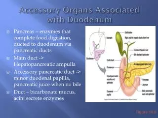
Mucous Membrane & Duodenal Papillae • The M.M.of 1st part of duodenum is smooth , but the remainder of duodenum , it is thrown into circular folds called the plicae circulares. • Halfwaydown the medial border of 2nd part of duodenum , the bile duct & the main pancreatic duct unite to form hepato-pancreatic ampulla that opens by major duodenal papilla.- the accessory pancreatic duct opens by a minor duodenal papilla above the major one.
Blood supply of Duodenum : • Arteries :upper ½ of duodenum … by superior pancreatico-duodenalartery , a branch of gastroduodenal lower ½ of duodenum … by inferior pancreatico-duodenalartery, a branch of superior mesenteric artery. • Veins :superior pancreatico-duodenal vein drains into the portal vein.Inferior pancreatico-duodenalvein drains into the superior mesenteric vein.
Lymph Drainage & innervation of the Duodenum : • It follows the arteries upward via pancreatico-duodenal nodes to gastro-duodenal L.Ns.and finally to celiac & hepatic L.Ns…. and downward via pancreatico-duodenal nodes to superior mesenteric L.Ns. • It is supplied by symp.& parasymp.vagal N.Fs.
Duodenal folds & Recesses : • They lie close to the duodeno-jejunaljunction, as 4 small pocketelike pouches of peritoneum. • Sup.duodenal fold of peritonium : on the left side of 4th part of duodenum, it has lower free border (recess). • Inf.duodenal fold of peritonium : on the left side of 4th part of duodenum, it has upper free border (recess).
Duodenal folds & recesses : • Paraduodenal fold & recess ofperitoneumlies to left of 4th part of duodenum. • It has free border which encloses the inferiormesenteric vein.
Duodenal folds & recesses : • The retroduodenalrecess is found behind 3rd &4th parts of duodenum. • The duodenum is connected to the posterior abdominal wall by a fold of peritoneum.
The Pancreas : • It is exocrine gland secrete enzymes for protein,fats & carbohydrates hydrolyzing. • It is endocrine gland that produce the hormonesinsulin & glucagon by pancreatic islets( islets of Langerhans) • It lies in epigastrium & left upperquadrant, crosses transpyloric plane (L1 vertebra). • It lies on the post.abd.wall behind the peritoneum ( retroperitoneal ),
Parts of Pancreas : • The head lies in the concavity of duodenum. • The uncinate process arises from the lower part of the head and lies behindsup.mesentericvessels. • The neck lies in front of the beginning of portal vein. • The body. • The tail passes in the splenico-renal ligament to end in the hilum of spleen.
The Relation of Pancreas : • Anteriorly : from right to left…. Transverse colon & mesocolon ,lesser sac & stomach. • Posteriorly : from right to left…. Bile duct, portal vein,superior mesenteric vessels,splenic vein -- I.V.C.,aorta. , Left psoas muscle.Left kidney., Left suprarenal and hilum of spleen.
Pancreatic Ducts : • The main pancreatic duct begins in the tail of pancreas and opens into middle of 2nd part ofduodenum by major duodenal papilla after joining the bile duct or drains separetely into the duodenum. • The accessory duct : when present , drains the upper part of head and opens into the duodenum by minor duodenalpapilla , a short distance above themain duct, frequently connects with the main duct.
Blood supply ,lymph drainage and innervation of Pancreas : • the arteries are :Splenic artery , superior & inferiorpancreatico-duodenal arteries. • The veins as arteries, drain into the portal system. • Lymph nodes lie along the arteries and drain into celiac & sup. mesenteric L.Ns. • Nerve supply : by symp.& parasymp.(vagal) N.Fs.
Cancer of head of pancreas leads to …… obstructive jaundice due to close relation of the head of pancreas to the bile duct. • Dudenal ulcer :at anterior wall of first inch ofduodenumdue to releasing acide chyme of stomach, may perforate into greater sac, above transverse colon which directs the escaping fluid into right iliac fossa,and confuse with perforated appendix. Ulcer of posterior wall of 1st part of duodenummay perforate thewall and erode gastrodudenal artery,causing a severe hemorrhage. • During Splenectomy , sometimes results in damage of the tail of pancreas which lies in the splenico-renal ligament , so the damaged pancreas releases enzymes that digest the surrounding tissues, leading to acute peritonitis.
Transpyloric plane (L1 vertebra) passes through : • Pylorus of stomach. • 1st part of duodenum. • Duodenojejunal junction. • Hilum of kidney. • Neck of pancreas. • Beginning of portal vein. • Abdominal aorta, at origin of sup.mesenteric artery. • Fundus of gall bladder (at tip of right 9th costal cartilage). • Opening of lesser sac.
Longitudinal section of2nd part of duodenum :showing the hepato-pancreaticampulla (ampulla of Vater) which is surrounded by sphincter of Oddi. • Note that the hepato-pancreatic ampulla is formed by union of bile duct & main pancreatic duct that opens into lumen of duodenum by majorduodenal papilla.