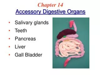
Accessory Organs Associated with Duodenum
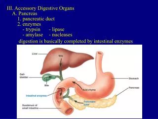
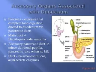
Accessory Organs Associated with Duodenum. Pancreas – enzymes that complete food digestion, ducted to duodenum via pancreatic ducts Main duct -> Hepatopancreatic ampulla Accessory pancreatic duct -> minor duodenal papilla, pancreatic juice when no bile

Accessory Organs Associated with Duodenum
E N D
Presentation Transcript
Accessory Organs Associated with Duodenum • Pancreas – enzymes that complete food digestion, ducted to duodenum via pancreatic ducts • Main duct -> Hepatopancreatic ampulla • Accessory pancreatic duct -> minor duodenal papilla, pancreatic juice when no bile • Duct – bicarbonate mucus, acini secrete enzymes Figure 14.6
Enzymes of Pancreas • Trypsinogen, chymotrypsinogen, procarboxypeptidase • Trypsinogen converted to Trypsin by enterokinase in lumen of duodenum • Trypsin cleaves other two to make chymotrypsin and carboxypeptidase, and digests dietary protein • Pancreatic amylase, pancreatic lipase, ribonuclease, deoxyribonuclease – active after exposure to bile and ions in lumen
Regulation of Pancreatic Secretions • Bile and pancreatic juice secreted in response to vagus stimulation (parasympathetic), inhibited by sympathetic • Both stimulated by Cholecystokinin (CCK), gastrin, and secretin • CCK released by duodenum in response to fat and acid from stomach. • Contraction of gallbladder – bile into duct • Secretion of pancreatic enzymes • Relaxation of hepatopancreatic sphincter • Secretin – stimulates bile and pancreatic duct secretion of bicarbonate, neutralize acid
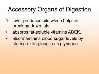
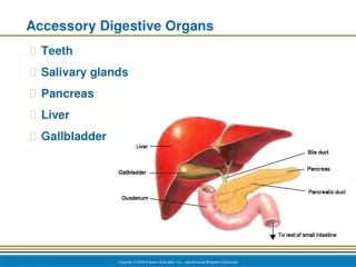
Liver • 4 lobes – rt, lt, quadrate, caudate, sep by falciform ligament = mesentary • Round ligament = remnant of umbilical vein, blod from umbilical cord to liver of fetus • Portal hepatitis – entry of hepatic portal vein, proper hepatic artery, exit of bile passages • Gallbladder associated
Microscopic liver • Hepatic lobules – central veins, cuboidal hepatocytes • Hepatic Triad – artery + vein (branches of proper hepatic artery and hepatic portal vein), bile ductule • Hepatic sinusoid – fenestrated endothelium sep hepatocyte from bloodstream, blood plasma fills, blood directly from intestine • Liver gets first dibs on glucose, AA, iron, vitamis, nutrients • Removes hormones, toxins, bile pigments, drugs • Secretes some blood proteins
Bile Secretion • Bile canaliculi, bile ductules of triads, left and right hepatic ducts ->common hepatic duct • Hepatic and cystic duct join = bile duct • Bile duct joins pancreatic duct = hepatopancreatic ampulla • Enter duodenum=major duodenal papilla, heptopancreatic sphincter (sp of Oddi)
Bile • Yellow-green color • Watery solution contain bile salts and pigments, cholesterol, phospholipids, electrolytes • Bilirubin – breakdown product of Hb • Only bile salts and phospholipids aid digestion – emulsify fats, more SA for fat digesting enzymes to work on
Accessory Digestive OrgansGall Bladder • Thin walled green sac • When not eating bile stored here • Concentrated by water removal • Fatty meal enters duodenum, bile released due to hormonal stimulus
Gallstones • Bile stored for too long or too much water removed, cholesterol crystallizes • Blockage of common hepatic or bile ducts prevent bile into small intestine, backs up into liver • Bile pigments enter blood and circulate, yellowing jaundice http://gogogojiteam.com/gallstone.html
Organs of Alimentary CanalSmall Intestine • MAJOR digestive organ • 3 subdivision • Duodenum • Jejunum • Ileum – meets large intestine at ileocecal valve • Only able to process small amount of food at time, pyloric sphincter controls food movement
Walls of Small Intestine • Nearly all food absorption • 3 structures in wall increase absorptive surface • Microvilli – plasma membrane – brush border enzymes – digestion of protein and carbs • Villi – mucosa, w/in each are capillaries and lacteal duct (fat), food absorbed, absorptive and goblet cells • Circular folds – deep fold of mucosa and submucosa, don’t disappear as fill with food • All decrease as move along • Peyer’s Patches increase – collections of lymphatic tissue • Bacteria in food! Figure 14.7
Cells of Villi • Absorptive cells line villi, have microvilli • Contain brush border enzymes – enterokinase – remember pancreatic Trypsinogen
Structures of Sm. Intestine • Crypts of Lieberkuhn – glands at base of villi, extend to muscularis mucosa – absorptive, goblet cells, stem cells, Paneth cells (lysozyme, phospholipase, defensins) • Brunner glands – submucosa – bicarbonate mucus (unique to duodenum) • Peyer patches – lymphatic nodules - ileum
Motility of Sm. Intestine • Segmentation vs. Peristalsis • Mix nutrients • Churn chyme, contact with mucosa for contact digestion and absorption • Move to large intestine
Organs of Alimentary CanalLarge Intestine • Ileocecal valve to anus, appendix • Absorb water and eliminate indigestible food as feces • No villi • Goblet cells in mucosa produce HCO-3 rich mucus, lubricant • Divisions • Cecum, Appendix, Colon, Rectum, Anal Canal • Colon – ascending, transverse, descending, sigmoid • Anus – internal and external sphincters, anal columns and sinuses Figure 14.8
Bacteria and Gas • Bacterial flora – ferment cellulose and other undigested nutrients • Synthesize B vitamins, vitamin K which are absorbed by colon • Don’t get enough vitamin K in diet • Expel about 500ml of gas • Some from swallowed air, some from bacterial digestion
Absorption • 12-24 from meal to feces • Reabsorbs water and electrolytes • Feces – 75% water and 25% solids • Solids – 30% bacteria, 30% undigested fiber, 10-20% fat (not diet), epithelial cells, salts, mucus, etc
Defecation • Stretching of rectum stimulates defecation • Intrinsic defecation reflex – stretch signals travel myenteric nerve plexus to muscularis of descending, sigmoid colon, rectum • Stimulates peristaltic waves • Internal sphincter relaxes – defecation if external also relaxed • Need cooperation of parasympathetic defecation reflex – spinal cord • Stretch of rectum, signal to sacral of spinal cord • Return via parasympathetic fibers, intensifies peristalsis • Parasympathetic fibers relax internal sphincter
Functions of the Digestive SystemIngestion • Food must enter the system to be acted upon • Active and voluntary Figure 14.11
Functions of the Digestive SystemPropulsion • Foods must be processed my multiple organs – move from one to next • Swallowing is example • Movement depends mostly on peristalsis vs. segmentation • Segmentation – small intestine, alternating segments contract, mechanical digestion
Functions of the Digestive SystemFood Breakdown: mechanical digestion • Mixing of food in mouth by tongue • Churning of food in stomach • Segmentation in small intestine • Prepare food for enzymatic digestion by breaking into smaller pieces
Functions of the Digestive SystemFood Breakdown: chemical digestion • Food molecules broken down into building blocks by enzymes • Reactions called hydrolysis rxn, water added as bond breaks • Carbs (saccharides) we digest – sucrose, lactose, maltose, and starch. Eat cellulose but can’t digest, fiber • Proteins (a.a.) – polypeptides or peptides • Lipids (fats) – fatty acids and glycerol
Functions of the Digestive SystemAbsorption • Transport of digested food into blood from lumen of GI tract • Must enter mucosal cells by active or passive transport. • Small intestine major site of absorption
Functions of the Digestive SystemDefecation • Elimination of indigestible food from GI tract via anus in form of feces
Digestion Review Figure 14.13 (1 of 3