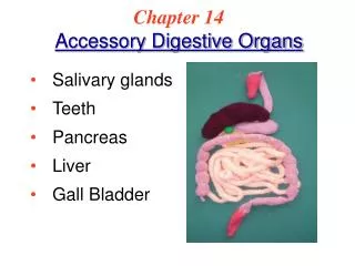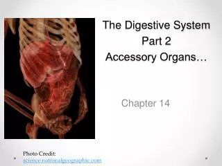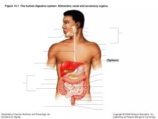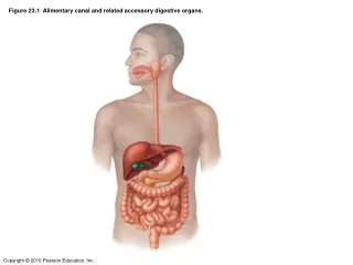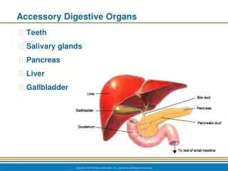
Accessory Digestive Organs
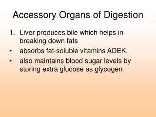
Accessory Digestive Organs. Salivary glands Pancreas Liver Gallbladder. Salivary Glands. Function: produce saliva Wets and lubricates the oral mucosa and the ingested food (→ bolus ) Initiates digestion of carbohydrates and lipids ( amylase and lingual lipase ). Salivary Glands.

Accessory Digestive Organs
E N D
Presentation Transcript
Accessory Digestive Organs • Salivary glands • Pancreas • Liver • Gallbladder
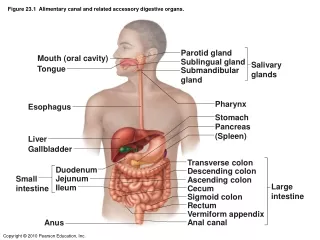
Salivary Glands • Function: produce saliva • Wets and lubricates the oral mucosa and the ingested food (→ bolus) • Initiates digestion of carbohydrates and lipids (amylase and lingual lipase)
Salivary Glands • Minor salivary glands • 10% of total volume of saliva, 70% mucus • Major salivary glands • Parotid • Submandibular (submaxillary) • Sublingual
Ductal System • Lobes and lobules separated by connective tissue septa • Secretory alveoli + ducts • Secretory alveoli: round, sac-like structures, simple cuboidal epithelium resting on a basal lamina • Ducts: intralobular, interlobular, lobar, main excretory ducts
Ductal System • Intralobular ducts – located within the lobule • 2 segments: • Intercalated duct – directly drains an acinus/secretory lobule • Striated (secretory) duct – union of intercalated ducts
Ductal System • Intercalated duct • Simple squamous or simple cuboidal epithelium • Contain myoepithelial cells • Striated duct • Simple cuboidal or simple columnar epithelium • Exhibit basal striations • Contain myoepithelial cells
Ductal System • Interlobular ducts – union of striated ducts, located in the connective tissue septa (between lobules) • Stratified cuboidal → stratified columnar • Lobar ducts – drain an entire lobe • Stratified columnar epithelium • Main excretory duct – opens into the oral cavity • Stratified squamous epithelium
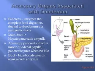
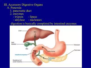
Pancreas • Pinkish, soft organ • 12-15 cm • Stretched transversely across the posterior abdominal wall from the duodenum to the spleen • Posterior to stomach • Mostly retroperitoneal
Pancreas • Head • Tail • Body • Main pancreatic duct (duct of Wirsung) • Accessory pancreatic duct (duct of Santorini)
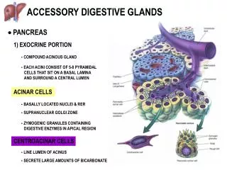
Pancreas • Main functions: • Exocrine: produces digestive enzymes that act in the small intestine (cells arranged in acini) • Endocrine: synthesizes and secretes hormones (insulin and glucagon) into the bloodstream (islets of Langerhans)
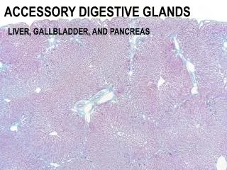
Exocrine Pancreas • Thin connective tissue septa divide the pancreas into lobules • Acini surrounded by a basal lamina that is supported by a delicate sheath of reticular fibers • Has rich capillary network (essential for the secretory process)
Pancreatic Acini • 40-50 closely packed low columnar or pyramidal cells • Narrow lumen, rest on basal lamina • Nucleus: round with 1 or more nucleoli • Cytoplasm: • Supranuclear – eosinophilic granules (zymogen granules) • Infranuclear (basal) – abundant rough ER
Pancreas vs. Parotid • Pancreas: • Absence of striated ducts • Presence of the islets of Langerhans • Initial portions of intercalated ducts penetrate the lumens of the acini (centroacinar cells – constitute the intra-acinar portion of the intercalated duct