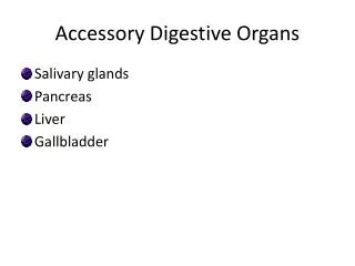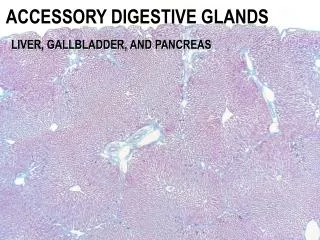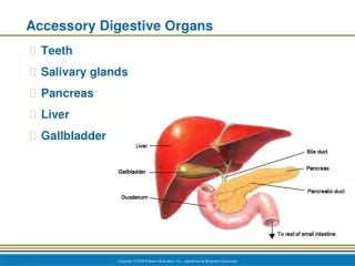
ACCESSORY DIGESTIVE GLANDS
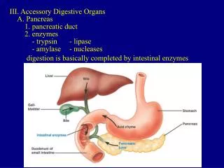
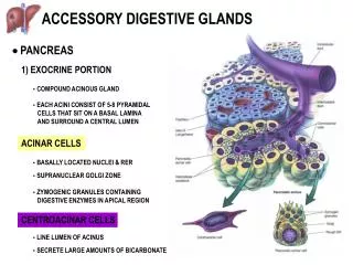
ACINAR CELLS. - BASALLY LOCATED NUCLEI & RER. - SUPRANUCLEAR GOLGI ZONE. - ZYMOGENIC GRANULES CONTAINING DIGESTIVE ENZYMES IN APICAL REGION. CENTROACINAR CELLS. - LINE LUMEN OF ACINUS. - SECRETE LARGE AMOUNTS OF BICARBONATE. ACCESSORY DIGESTIVE GLANDS . PANCREAS.

ACCESSORY DIGESTIVE GLANDS
E N D
Presentation Transcript
ACINAR CELLS - BASALLY LOCATED NUCLEI & RER - SUPRANUCLEAR GOLGI ZONE - ZYMOGENIC GRANULES CONTAINING DIGESTIVE ENZYMES IN APICAL REGION CENTROACINAR CELLS - LINE LUMEN OF ACINUS - SECRETE LARGE AMOUNTS OF BICARBONATE ACCESSORY DIGESTIVE GLANDS PANCREAS 1) EXOCRINE PORTION - COMPOUND ACINOUS GLAND - EACH ACINI CONSIST OF 5-8 PYRAMIDAL CELLS THAT SIT ON A BASAL LAMINA AND SURROUND A CENTRAL LUMEN
ACCESSORY DIGESTIVE GLANDS PANCREAS 1) EXOCRINE PORTION INTERCALATED DUCTS PANCREAS H&E
ACCESSORY DIGESTIVE GLANDS PANCREAS 2) ENDOCRINE PORTION
ACCESSORY DIGESTIVE GLANDS LIVER PORTA HEPATIS PORTAL TRIAD
- central vein at center - hexagonal in shape - short axis: branches of portal triad between 2 classic lobules - portal triad at corners - long axis: between 2 central veins - portal triad at center - triangular in shape - central vein at corners ACCESSORY DIGESTIVE GLANDS LIVER CLASSIC LOBULE PORTAL LOBULE LIVER ACINUS
ACCESSORY DIGESTIVE GLANDS LIVER
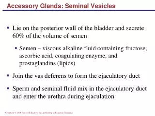
HEPATOCYTE BILE CANALICULI RT & LT HEPATIC DUCTS COMMON BILE DUCT ACCESSORY DIGESTIVE GLANDS GALLBLADDER - LOCATION FOR CONCENTRATION AND STORAGE OF BILE ROUTE OF BILE FILLING OF GALLBLADDER
URINARY SYSTEM: I TO IDENTIFY THE COMPONENTS OF THE URINARY SYSTEM TO CHARACTERIZE THE GENERAL ORGANIZATION OF THE KIDNEY TO EXAMINE THE HISTOLOGICAL STRUCTURE OF THE NEPHRON AND THE COLLECTING DUCTS TO CORRELATE STRUCTURE OF VARIOUS COMPONENTS WITH FUNCTION
URINARY SYSTEM KIDNEY ANATOMICAL STRUCTURE AND BLOOD SUPPLY - highly vascular (25% cardiac output) - produces urine (water and elctrolytes, urea, uric acid, creatinine), initially an ultrafiltrate of the blood URETER BLADDER URETHRA
URINARY SYSTEM KIDNEY 1) EXOCRINE PORTION 2) ENDOCRINE PORTION - synthesis and secretion of erythropoietin (regulation of red blood cell formation) - synthesis and secretion of renin (hormone necessary for control of blood pressure and blood volume)
URINARY SYSTEM KIDNEY (ORGANIZATION) GROSS STRUCTURE: - RENAL HILUM, PELVIS, AND SINUS C - RENAL CAPSULE M - RENAL CORTEX - RENAL MEDULLA
P P P P P RC P P URINARY SYSTEM KIDNEY (ORGANIZATION) CORTEX - region immediately beneath renal capsule - composed of two distinct regions: (1) CORTICAL LABYRINTH (2) MEDULLARY RAY MEDULLA - located immediately beneath renal cortex - consists of triangular blocks of tissue called the PYRAMIDS - RENAL COLUMNS are strands of cortical tissue that extend down between adjacent pyramids
Cortical Labyrinth with interdigitating Medullary Rays RENAL LOBE - a single pyramid with its associated overlying cortex RENAL LOBULE - defined within cortex and involves a single medullary ray (central axis of lobule) with adjacent adjacent cortical labyrinth - defined as a functional unit that consists of a collecting duct and all the nephrons that it drains URINARY SYSTEM KIDNEY (ORGANIZATION) P P P P P P P
URINARY SYSTEM THE NEPHRON & COLLECTING DUCTS
URINARY SYSTEM THE NEPHRON & COLLECTING DUCTS 1) THE NEPHRON - distributed throughout cortex and various zones of medulla a) RENAL CORPUSCLE BOWMAN’S CAPSULE + GLOMERULUS b) PROXIMAL TUBULE CONVOLUTED AND STRAIGHT PORTIONS c) HENLE’S LOOP THICK AND THIN PORTIONS d) DISTAL TUBULE STRAIGHT AND CONVOLUTED PORTIONS 2) COLLECTING DUCTS
URINARY SYSTEM THE NEPHRON & COLLECTING DUCTS CORTEX: CORTICAL LABYRINTH 1- RENAL CORPUSCLES 2- PROXIMAL CONVOLUTED TUBULES 3- DISTAL CONVOLUTED TUBULES MEDULLARY RAY 1- STRAIGHT PORTIONS OF PROXIMAL TUBULE (THICK DESCENDING) 2- STRAIGHT PORTIONS OF DISTAL TUBULE (THICK ASCENDING) 3- COLLECTING DUCTS
URINARY SYSTEM THE NEPHRON & COLLECTING DUCTS MEDULLA: OUTER ZONE 1- STRAIGHT PORTIONS OF PROXIMAL TUBULE (THICK DESCENDING) 2- STRAIGHT PORTIONS OF DISTAL TUBULE (THICK ASCENDING) 3- THIN SEGMENTS OF LOOP OF HENLE (DESCENDING & ASCENDING) 4- COLLECTING DUCTS INNER ZONE 1- THIN SEGMENTS OF LOOP OF HENLE (DESCENDING & ASCENDING) 2- COLLECTING DUCTS
RENAL LOBULE URINARY SYSTEM BLOOD FLOW (KIDNEY) AORTA RENAL ARTERY INTERLOBAR ARTERIES - run between lobes in medulla ARCUATE ARTERIES - run parallel to bases of pyramids at the corticomedullary junction INTERLOBULAR ARTERIES - delineate lateral limits of renal lobules AFFERENT ARTERIOLES - supply blood to glomerulus GLOMERULAR CAPILLARY BED EFFERENT ARTERIOLES - drain blood from glomerulus and form either peritubular capillary plexus (cortex) or vasa recta system (medulla)
RENAL LOBULE URINARY SYSTEM BLOOD FLOW (KIDNEY) VENA CAVA RENAL VEIN INTERLOBAR VEINS - run between lobes in medulla ARCUATE VEINS - run parallel to bases of pyramids at the corticomedullary junction INTERLOBULAR VEINS - delineate lateral limits of renal lobules PERITUBULAR CAPILLARY PLEXUS VASA RECTA SYSTEM
URINARY SYSTEM ea G G BLOOD FLOW (KIDNEY) aa G IA
URINARY SYSTEM THE NEPHRON & COLLECTING DUCTS HISTOLOGICAL STRUCTURE AND FUNCTION 1) THE NEPHRON - distributed throughout cortex and various zones of medulla a) RENAL CORPUSCLE BOWMAN’S CAPSULE + GLOMERULUS b) PROXIMAL TUBULE CONVOLUTED AND STRAIGHT PORTIONS c) HENLE’S LOOP THICK AND THIN PORTIONS d) DISTAL TUBULE STRAIGHT AND CONVOLUTED PORTIONS 2) COLLECTING DUCTS
URINARY SYSTEM THE NEPHRON & COLLECTING DUCTS HISTOLOGICAL STRUCTURE AND FUNCTION 1) THE NEPHRON - distributed throughout cortex and various zones of medulla a) RENAL CORPUSCLE BOWMAN’S CAPSULE + GLOMERULUS b) PROXIMAL TUBULE CONVOLUTED AND STRAIGHT PORTIONS c) HENLE’S LOOP THICK AND THIN PORTIONS d) DISTAL TUBULE STRAIGHT AND CONVOLUTED PORTIONS 2) COLLECTING DUCTS
URINARY SYSTEM RENAL CORPUSCLE BOWMAN’S CAPSULE + GLOMERULUS FILTRATION APPARATUS OF KIDNEY 1. BOWMAN’S CAPSULE: - the beginning of the nephron that consists of a blind sac lined with simple squamous epithelium that is continuous with the PCT - parietal layer & visceral layer (specialized) 2. GLOMERULUS: - specialized tuft of capillaries which housed in the capsular space (10-20 capillary loops) - blood flowing through glomerulus capillaries undergoes a filtration process to produce the initial urine filtrate
URINARY SYSTEM RENAL CORPUSCLE BOWMAN’S CAPSULE + GLOMERULUS FILTRATION APPARATUS OF KIDNEY VASCULAR POLE URINARY POLE GLOMERULUS (FILTRATION MEMBRANE): 1- fenestrated capillaries; discontinuous endothelium; fenestrae have a diameter of 500-1000Å and lack a diaphragm 2- continuous basal lamina 3- podocytes of visceral layer; processes contact basal lamina and are separated by slits measuring approximately 250Å
URINARY SYSTEM RENAL CORPUSCLE BOWMAN’S CAPSULE + GLOMERULUS FILTRATION APPARATUS OF KIDNEY GLOMERULUS (FILTRATION MEMBRANE): prevents RBC’s and large MW proteins from leaving circulation, while most other blood constituents pass easily into the capsular space MESANGIAL CELLS - phagocytic cells with a surrounding matrix that lend structural support to the glomerulus
URINARY SYSTEM RENAL CORPUSCLE BOWMAN’S CAPSULE + GLOMERULUS FILTRATION APPARATUS OF KIDNEY GLOMERULUS (FILTRATION MEMBRANE): 1- fenestrated capillaries 2- continuous basal lamina 3- podocytes
PODOCYTE 1° process pedicels 2°