Download
1 / 30
300 likes | 590 Vues
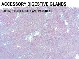
Large glands associated with the digestive tract. Salivary glands 2 main groups 1. Major salivary glands: paired parotid Submandibular Sublingual 2. Minor salivary glands : Buried in the C/T of lips, cheeks, tongue, palate (produce 5% of total salivary output).

E N D
Salivary glands • 2 main groups • 1. Major salivary glands: • paired parotid • Submandibular • Sublingual • 2. Minor salivary glands : • Buried in the C/T of lips, cheeks, tongue, palate (produce 5% of total salivary output)
The secretion of each gland is either serous, seromucous, or mucous, depending on its glycoprotein mucin content. • Saliva from the parotids is serous and watery. • The submandibular and sublingual glands produce a seromucous secretion, with mostly mucus from the minor glands.
The main functions of the salivary glands • to wet and lubricate ingested food and the oral mucosa, • to initiate the digestion of carbohydrates and lipids with amylase and lipase, • and to secrete protective bacteriostatic substances such as the immunoglobulin IgA, lysozyme, and lactoferrin.
In the large salivary glands, the connective tissue contains many lymphocytes and plasma cells. • The plasma cells release IgA, which forms a complex with a secretory component synthesized by the epithelial cells of serous acini and intralobular ducts. • The IgA-secretory complex released into the saliva resists enzymatic digestion and constitutes an immunologic defense mechanism against pathogens in the oral cavity.
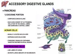
Salivary glands • Compound tubuloacinar glands • Ducts open in the oral cavity • Surrounded by a C/T capsule • From capsule, C/T septa extend into the gland and divide it into lobe and lobules. • Blood vessels, lymphatics, nerves, excretory duct of glands
Secretoryacini are surrounded and supported by very fine loose C/T • Lymphocytes, plasma cells. • C/T of acini blends C/T of septa
Parenchyma of salivary glands • Functional glands • Composed of acini (alveoli) • Some portions of ducts are involved in secretory process so called as tubuloacinar glands. • Acini are spherical structures consisting of secretory epithelial cells arranged around a central cavity which continues into that of the duct draining the acinus. • The acini of salivary glands contain serous cells, mucous cells or both.
Serous acini • Serous acini are generally spherical structures, the mucous end pieces are more often tubular. • Pyramidal in shape • With a broad base facing the basal lamina • Narrow apical surface facing the lumen of the acinus. • Basally located spherical nucleus.
Basal region of cell contains numerous mitochondria, RER, free ribosomes • Apex contains zymogenic granules • well developed GA is present between nucleus and secretory granules • Basal region (basophilic due to ribosomes • Apical region (acidophillic due to zymogenic granules)
Adjacent serous cells are joined to each other by junctional complexes • apical to junctional complexes intercellular canaliculi exist between the adjacent serous cells • Serous cells produce thin watery secretion which is rich in digestive enzymes.
Mucous cells • Pyramidal in shape • Nuclei are oval (or flattened) and pressed against the basal plasmalemma • Apical region of each mucous cells is occupied by numerous mucinogen granules , stained poorly and appear to be empty
Mucous cells contain mitochondria, moderate amount of RER, very large golgi apparatus(which add carbohydrates to proteins to produce glycoproteins of mucinogen which is a precursor of mucin). • Adjacent cells are bound together by apical junctional complexes and do not exhibit intercellular canaliculi.
The mucous cells secrete mucin which mixes with water and coverted to mucus which is a gel like lubricant- protect the delicate mucous membrane. • Mucous cells are most often organized as tubules rather than acini and produce mostly mucins.
Myoepithelial cells, • are found inside the basal lamina of the secretoryunits • and (to a lesser extent) the initial part of the duct system • Surrounding the secretory portion myoepithelial cells are well developed and branched (and are sometimes called basket cells), whereas those associated with the initial ducts are spindle-shaped and lie parallel to the duct's length.
Myoepithelial cells prevent distention of the endpiece when the lumen fills with saliva and their contraction accelerates secretion of the product. • Has a body and many long processes • Cell body contains nucleus and small amount of organelle • Cell process contain actin and myosin. • Stained poorly and cannot be easily identified.
Duct system of salivary glands • Compound salivary glands have highly branched duct system • 3 types • 1. intercalated ducts intralobular ducts • 2. striated ducts • 3. excretory ducts
Intercalated ducts • In the intralobular duct system,secretoryendpieces empty into intercalated ducts, lined by cuboidal epithelial cells, • Have small diametre • Cuboidal epithelial cells have capability to divide and differentiate into secretory cells and ductal cells • These cells also secrete carbonic anhydrase (secrete bicarbonate and absorb chloride ions from it) • and several of these short ducts join to form striated ducts • Intercalated ducts in parotid and submandibular are long • Very short in sublingual
Striated ducts • The columnar cells of striated ducts often show radial striations extending from the cell bases to the level of the nuclei. • Larger caliber • Stain intensely with eosin • Nucleus is central • Due to presence of prominent eosinophillic striations, these ducts are called striated ducts
Ultrastructurally the striations consist of infoldings of the basal plasma membrane • Numerous mitochondria are aligned parallel to the infolded membranes which contain ion transporters. • Such folds greatly increase the cell surface area, facilitating ion absorption, and are characteristic of cells specialized for ion transport.
Absorbtion of Na+ • Secretion of K+ so reduce the tonicity of saliva
Excretory ducts. • The striated ducts of each lobule converge and drain into ducts located in the connective tissue septa separating lobules, where they become interlobular, or excretory ducts. • Interlobular= pseudostratified or stratified cuboidal epithelium, • Interlobar=pseudo stratified columnar epithelium containing a few mucous cells. • The main duct = stratified columnar epithelium and then nonkeratinized-stratified squamous epithelium.
Interlobular ducts join to form interlobar ducts, which ultimately drain into main duct of the gland which drain secretory products of the gland into oral cavity • interlobular, interlobar and main ducts are purely secretory, i.e. they are simple conduits and donot modify the secretory product.
Vessels and nerves enter the large salivary glands at a hilum and gradually branch into the lobules. A rich vascular and nerve plexus surrounds the secretory and ductal components of each lobule. • The capillaries surrounding the secretory end pieces are very important for the secretion of saliva, which is stimulated by the autonomic nervous system. • Parasympathetic stimulation, usually elicited through the smell or taste of food, provokes a copious watery secretion with relatively little organic content. • Sympathetic stimulation inhibits such secretion, and produces the potential for dry mouth often associated with anxiety.