Comprehensive Guide to Digestive Tract Anatomy | Understanding the GI System
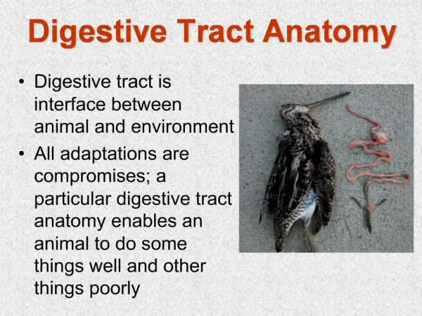
Explore the anatomy of the digestive tract, from the oral cavity to the small and large intestine. Learn about the structures like teeth, tongue, salivary glands, and stomach. Understand the innervation and blood supply of the GI system.

Comprehensive Guide to Digestive Tract Anatomy | Understanding the GI System
E N D
Presentation Transcript
Digestive tract Dr. Szuák András GYTK 2018.03.05.
Parts of the digestive tract • Oral cavity • Teeth • Tongue • Salivary glands • Pharynx • Esophagus • Stomach • Intestine • Small intestine • Large intestine
Oral cavity – cavum oris • Parts: • Oral vestibule (1) • Oral cavity proper (2) • Isthmus of fauces (3)
Teeth • Deciduous teeth • 2 incisor teeth • 1 canine tooth • 0 premolar tooth • 2 molar teeth • Permanent teeth • 2 incisor teeth • 1 canine tooth • 2 premolar teeth • 3 molar teeth
Teeth • Crown • enamel cover • Neck • meets with gingiva • Root • cementum cover
Tooth development dentin predentin odontoblasts in dental papilla dentin enamel rods ameloblasts (inner enamel epithelium)
Tongue root terminal sulcus palatine tonsil palatine tonsil lingual tonsil circumvallated papillae foliatae papillae apex fungiform papillae filiform papillae caruncula
Tongue Many papillae on the surface • Filiform papillae • Fungiform papillae • Foliate papillae • Circumvallate papillae • Taste buds
fungiform papilla filiform papilla
Papillae Fungiform papilla Circumvallate papilla Foliate papillae
Innervation of the tongue Sensory innervation: general sensory: lingual n. (V./3) – anterior 2/3 glossopharyngal n. (IX.) – posterior 1/3 vagus n. (X.) taste: chorda tympani (VII.) – anterior 2/3 glossopharyngal n. (IX.) – posterior 1/3 vagus n. (X.) tounge near epiglottis Motor innervation: hypoglossal n. (XII.)
Salivary glands • Submandibular gl. • Sublingual gl. • Parotid gl. • Small glands • Labial gl. • Buccal gl. • Palatine gl. • Lingual gl.
Salivary glands submandibular sublingual parotid
Isthmus of fauces palatoglossal arch uvula palato- pharyngeal arch * * *: palatine tonsil tongue
Pharynx Parts: • Nasopharynx / Epipharynx • Nasal cavity (choana) • Middle ear (tuba auditiva) • Oropharynx / Mesopharynx • Oral cavity (isthmus of fauces) • Laryngeopharynx / Hypopharynx • Larynx (laryngeal inlet) Innervation: glossopharyngeal n. (IX), vagus n. (X)
Pharynx Nasal cavity (choana) Oral cavity (isthmus of fauces) Laryngeal inlet
Esophagus • Continuation of pharynx • Passes through the thorax • Passes through the diaphragm • Reaches the stomach • Histology of esophagus… • but first, it’s neccessary to understand the general structure of the wall in the digestive tract
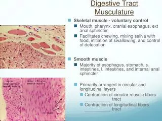
Structure of the wall in the GI tract • Mucosa • Epithelium (1) • Lamina propria (2) • Muscularis mucosae (3) • Submucosa (4) (pl. submucosus – Meisner’s) • Muscularis externa (pl. myentericus – Auerbach’s) • Inner circular (5) • Outer longitudinal (6) • Adventitia / Serosa (7)
Esophagus • Non-keratinized stratified squamous epith. • Cardia-glands in the lamina propria • Muscularis mucosae is longitudinal • Glandulae esophageae in submucosa • Muscularis externa • Adventitia in the thoracic cavity, serora in the abdominal cavity
Stomach Parts: • Fundus • Corpus • Antrum • Pylorus
Topography of the stomach Left coronary lig. Gastrophrenic lig. Gastrosplenic lig. Phrenicocolic lig. Hepatoduodenal lig. Lesser omentum Transverse mesocolon Greater omentum Inferior recess Diaphragm Spleen Stomach Liver Left colic flexure Right colic flexure Transverse colon Descending colon Ascending colon
Splenic a. Left gastric a. Short gastric aa. Right gastric a. Proper hepatic a. Gastroduodenal a. Left gastroomental a. Common hepatic a. Right gastroomental a. Blood supply of the stomach Celiac trunk Abdominal aorta
V. lienalis V. gastrica brevis V. gastrica sin. V. gastrica dext. Vena portae V. prepylorica V. gastroepiploica sin. V. mesenterica inf. V. pancreatico-duodenalis V. gastroepiploica dext. V. mesenterica sup.
Stomach • Simple columnar epith. Gastric pits • Lamina propria: long, tubular glands • Mucous secreting neck cells • Chief cells on the base – pepsinogen • Parietal cells – gastic acid • Muscularis mucosae • Inner circular&outer longitudinal layers • Submucosa • Muscularis externa • 3 layers: inner oblique, middle circular, outer longitudinal • Serosa
Smallintestine • Parts of theduodenum: • Superior part • Descending part • Horizontal part • Ascending part Jejunum: usually vertical Ileum: usually horizontal
Blood supply of the intestine - Superior mesenteric a. Superior mesenteric a. Inf. pancreaticoduodal a. Anastomosis with celiac trunk Anastomosis with inferior mesenteric a. Middle colic a. Right colic a. Jejunal aa. Ileocolic a. Ileal aa. Appendicular a.
Histology of the small intestine Structures helping absorbtion (with surface enlargment): • Circular folds(plicae circulares): More prominent in the duodenum and in the proximal part of the jejunum. They disappear at the terminal part of the ileum. • Villus (villi intestinales): projecting parts of the mucosa. They are more numerous in the duodenum and in the jejunum. Size: 0,5-1,5 mm • Microvilli: Projections on the apical surface of the epithelial cells. 3 1 2
Intestinal vili (Scanning Electron Microscopy) Lieberkühn cripts vili
Histology of the small intestine villi Lieberkühn cripts (intestinal galands) epithelium lamina propria Mucosa muscularis mucosae Submucosa circular longitudinali Muscularis externa Serosa
Paneth cells Duodenum Vili Lieberkühn cripts Muscularis mucosae Bruner’s glands Muscularis externa
Large intestine • Coecum • Vermiform appendix • Ascending colon • Transverse colon • Descending colon • Sigmoid colon • Rectum
Inferiormesenteric a. Abdominal aorta Anastomosis with superior mesenteric a. Left colic a. Middle colic a. Inferior mesenteric a. A Sigmoid aa. B Superior rectal a.
Colon • Simple coumnar epith. with many goblet cells • Lieberkühn cripts in the l. propria • Muscularis mucosae • Inner circular&outer longitudinal layers • Submucosa – NO glands • Muscularis externa • Inner circular&outer longitudinal layers the outer layer is not continous, it forms the 3 tenia coli • Serosa OR Adventitia
Appendix • Lymph nodules in the lamina propria and submucosa • Tenia coli is absent
Rectum • The veins form porto-caval anastomosis! Upper 1/3 v. portae Lower 2/3 v. cava inf.