Download
1 / 69
700 likes | 838 Vues
Learn about the essential function and process of the digestive system for breaking down food materials, absorption, and nutrient utilization. Explore the components of the digestive tract and the chemical digestion processes involved. Discover the structural characteristics of organs like the stomach and esophagus.

E N D
Digestive tract Li Zhong Jie(李仲杰), Ph. D School of Medicine, Zhejiang University lizhongjie@zju.edu.cn
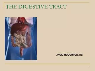
---Digestive system: • Digestive tract • Digestive gland ---Digestive system function: This system is responsible for the mechanical and chemical break down of food material, and for absorbing these digestive products into the blood for use as nutrients by the individual cells and tissues of the body
The Digestive Process • Ingestion • Taking in food through the mouth • Propulsion (movement of food) • Swallowing • Peristalsis – propulsion by alternate contraction & relaxation • Mechanical digestion • Chewing • Churning in stomach • Mixing by segmentation • Chemical digestion • Carbohydrates, Fat, and Proteins are broken down by enzymes. • Absorption • Transport of digested end products into blood and lymph in wall of canal • Defecation • Elimination of indigestible substances from body as feces
Chemical digestion --- Complex food molecules (carbohydrates, proteins and lipids) broken down into chemical building blocks (simple sugars, amino acids, and fatty acids and glycerol) --- Carried out by enzymes secreted by digestive glands into lumen of the alimentary canal
Components of digestive tract ---oral cavity ---pharynx ---esophagus ---stomach ---small intestine ---large intestine ---rectum and anus
General plan of digestive tract ---Except for oral cavity and pharynx, all other organs share a similar histological plan • Mucosa • Submucosa • Muscularis externa • Adventitia from lumen (inside) out
Inner layer: the mucosa*(mucous membrane) Three sub-layers • epithelium • lamina propria (may contain glands) • Muscularismucosae (Smooth muscle) *
Mucosa 1. Epithelium -------two types stratified squamous & simple columnar epith. 2. Lamina propria LCT. contained small glands,blood & lymph capillaries. mucosa-associated lymphoid tissue 3. Muscularis mucosa inner circular / outer longitudinal layer of smooth muscle cells. surface depth
the submucosa* • LCT. with small blood / lymphatic vessels; • *glands only in the • esophagus and • duodenum • Submucosal ( Meissner’s autonomic) nerve plexus *
Plica *a fold of mucosa and submucosa *longitudinal/circular form.
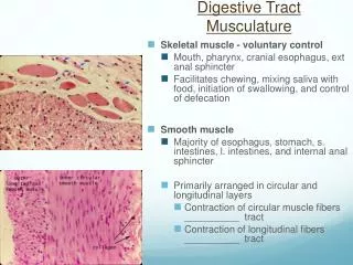
the muscularis externa* Two layers of smooth muscle responsible for peristalsis and segmentation • Inner circular layer (circumferential) • Myenteric (Auerbach’s autonomic) nerve plexus • Outer longitudinal layer: ---shortens gut *
Smooth muscle • Muscles are spindle-shaped cells • One central nucleus • Grouped into sheets: often running perpendicular to each other • Peristalsis • No striations (no sarcomeres) • Contractions are slow, sustained and resistant to fatigue • Does not always require a nervous signal: can be stimulated by stretching or hormones
Adventitia *the outmost layer formed by CT with two types: fibrosa: CT blending with surrounding structure serosa: CT + mesothelium (for example, peritoneum)
Enteric nervous system: • Mainly components: • 100 million neurons! (as many as the spinal cord) • unmyelinated nerve fibers • controlling the muscles, glands and having sensory info • Muscularis external: Myenteric nerve plexus • Submucosa: Submucosal nerve plexus
*Myenteric (Auerbach’s) plexus : regulate the movement of SM inner circular Nerve plexus outer longitudinal
Esophagus mucosa submucosa muscularis adventitia
Esophagus Passage way for food from the pharynx to the stomach mucosa: • epithelium: stratified squamous epithelium • lamina propria: compact CT contain simple tubular glands • muscularis mucosa: longitudinal arranged smooth muscle • At EG junction – thin simple columnar epithelium submucosa: • LCT • esophageal gland: mucous gland ---acidic mucin
Muscularis externa: • inner circular and outer longitudinal • upper 1/3: skeletal muscle • middle 1/3: mixed of skeletal muscle and smooth muscle • lower 1/3: smooth muscle Tunica adventitia: a fibrous coat of loose connective tissue
Cardiac Junction • Epithelial transition • ---Stratified Squamous nonkeratinized to simple columnar • It is clinically important, as it is the most site of esophageal carcinoma.
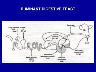
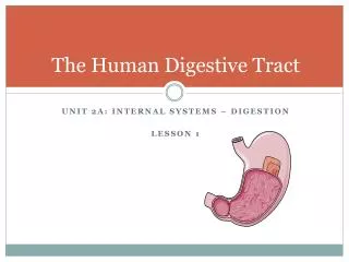
Stomach ---widest part of alimentary canal ---Temporary storage and mixing – 4 hours ---digest food partially to form a semi-fluid mass, termed chyme ---absorb part of water and ions • Water, electrolytes, some drugs like aspirin and alcohol (absorbed through stomach)
Stomach (regions) • Cardia • Surrounds esophageal entrance • Fundic stomach defined histologically includes • Fundus • Body • Pylorus • Pylorus is continuous with the duodenum
plica: longitudinal folds on internal surface (helps distensibility)
Structural Characteristic of Stomach • Mucosa • Epithelium (simple columnar mucus-secreting) • Lamina propria (gastric glands of different types) • Muscularismucosae (Smooth muscle) • Submucosa • Loose C.T. no glands • Muscularisexterna • inner oblique, middle circular, outer longitudinal • Tunica adventitia • Mostly serosa
mucosa • plica • Longitudinal folds of mucosa • A mucosal fold contains submucosa • Gastric pits: small depressions, 3-5 gastric gland open into the bottom • Diffuse lymphoid tissue and nodules may be present Gastric pit
plicain the stomach Mucosa Muscularis mucosa Submucosa Muscularis externa plica
①epithelium: simple columnar epithelium • surface mucous cell: ---tall columnar ---ovoid, basally-located nuclei ---apical mucin granule ---tight junction *gastric pits
Cross section of gastric pits Simple columnar epithelium Gastric pit Laminia propria between pits
②lamina propria: CT contains fibroblast, LC, plasma cell, mast cell and eosinophilic smooth muscle *Gastric gland: • fundic gland:oxyntic gland • cardiac gland: mucous gland • pyloric gland: mucous gland
* Fundic gland ---long, straight, branched or unbranched gland
Three part of gland: The neck The body The base Five type cells are found: Chief cells Parietal cells Mucous neck cells Stem cells endocrine cells
parietal cell or oxyntic cell ---structure: LM: • large, pyramidal or spherical • round centrally-located nuclei • eosinophilic cytoplasm
EM: • intracellular secretory canaliculus (active stage) • tubulovesicular system(resting stage) • a few RER, Golgi apparatus and more mitochondria secreting stage Resting stage
intracellular secretory canaliculus tubulovesicular system microvilli nucleus mitochondria Resting stage Secreting stage Ultrastructural model of Parietal cell
intracellular secretory canaliculus Secreting stage interconvert Resting stage tubulovesicular system
---function: i. secret hydrochloric acid (HCl) synthesis processes of HCl: in intracellular secretory canaliculus • H+ K+ -ATP pump: get H+ from cell • Cl- channel: get Cl- from blood • H+ +Cl-→HCl function of HCl: • pepsinogen→pepsin • kill the bacteria
ii.secret intrinsic factor: glycoprotein + Vitamin-B12→absorption of VB12 in ileum an deficiency of VB12 result in Addison’s anaemia
chief cell ( zymogenic cell) ---structure: LM: • columnar • Round, basally-located Nuclei • cytoplasm: /basal-basophilic /apical-zymogen granules
EM: • RER, Golgi apparatus ---function: secret pepsinogen(the precursor of pepsin)
Function of chief cell secreting pepsinogen (inactive) pepsin (active) protein--------peptides (acid environment)
主细胞 内分泌细胞 颈粘液细胞 壁细胞 Fundic gland
chief cell parietal cell secret hydrochloric acid secret intrinsic factor function: secret pepsinogen
mucous neck cell • less, neck part • pale stain in HE stain • secrete mucus stem cell undifferentiated cell endocrine cell • L cell: secreting histamine, promote secretion of parietal cell • D cell: secreting somatostatin, inhibit the secretion of parietal cell
* Mucous-HCO3barrier structure : • mostly of glycoproteins rich in carbohydrates and bicarbonate ions • 0.25-0.5mm thick mucous • The pH of the surface is usually 0.9-1.5
* Mucous-HCO3barrier function : • The mucus( bicarbonate-buffered mucus) is secreted on to the epithelial surface to form a barrier layer which protects it from injury by ingested substance and the stomach’s own secretion of acid and enzymes. • acid-base neutralization H+ + HCO3- → H2CO3 →H2O + CO2 ↑ bicarbonatase
4.Match the cell type with its secretion: 1. G-cell A. gastrin 2. Parietal Cell B. Bicarbonate ion 3. Chief Cell C. pepsinogen 4. Goblet Cell D. intrinsic factor 5. ECL Cell E. mucous 6. Surface mucus cell F. histamine
Small intestine • Duodenum – first region, only about 25cm long, • Jejunum – second region is roughly 2.5m long • Ileum – last region is roughly 3.5m long • Primary functions • Transport food from stomach to Large intestine • Secretion of digestive enzymes to facilitate digestion of food substances • Absorption of nutrient substances into blood and lymph vessels • Secretion of certain hormones
Special structure of mucosa • Plicae circulares • Mucosa and submucosa are arranged in permanent, circular mucosal folds • Intestinal villi • Mucosal projections covered by epithelium and containing only lamina propria • Crypt or intestinal glands • Surrounded by lamina propria • Extend to the muscularis mucosae
villi Plicaecirculares