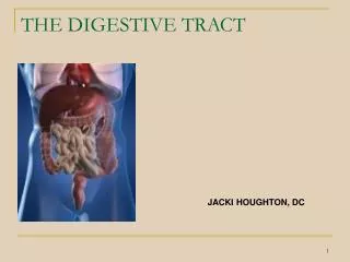
Digestive tract diseases
Digestive tract diseases. Adam Stefański. Esophagitis - causes. gastroesophageal reflux infectious esophagitis (in patients who are immunocompromised) radiation direct erosive effects of ingested medication or corrosive agents. Pathophysiology of esophagitis.


Digestive tract diseases
E N D
Presentation Transcript
Digestive tract diseases Adam Stefański
Esophagitis - causes • gastroesophageal reflux • infectious esophagitis (in patients who are immunocompromised) • radiation • direct erosive effects of ingested medication or corrosive agents.
Pathophysiology of esophagitis Reflux esophagitis develops when gastric contents are passively regurgitated into the esophagus. Gastric acid, pepsin, and bile irritate the squamous epithelium, leading to erosion and ulceration of esophageal mucosa. Eventually, a columnar epithelial lining may develop. This lining is a premalignant condition termed Barrett esophagus.
Esophagitis - medical history • The most common complaint is heartburn (dyspepsia), a burning sensation in the mid chest caused by contact of stomach acid with inflamed esophageal mucosa. Symptoms often are maximal while supine, when bending over, when wearing tight clothing, or after large meals • Water brash is a bitter taste of refluxed gastric contents often associated with heartburn • Other common symptoms include upper abdominal discomfort, nausea, bloating, and fullness. Less common symptoms include dysphagia, odynophagia, cough, hoarseness, wheezing, and hematemesis
Esophagitis - medical history • The patient may experience chest pain indistinguishable from that of coronary artery disease. Pain often is midsternal with radiation to the neck or arm and may be associated with shortness of breath and diaphoresis. Chest pain may be relieved with nitrates if esophageal spasm is involved, further confounding diagnostic evaluation. • Infants with reflux are at greater risk of aspiration. Symptoms include weight loss, regurgitation, excessive crying, back arching, respiratory distress, and apnea.
Esophagitis – physical examination • Physical examination usually is not helpful in uncomplicated esophagitis, but evaluate other potential sources of chest pain such as the chest wall and abdomen. • Perform a rectal examination for occult bleeding.
Conditions that may increase risk of reflux esophagitis • Pregnancy • Obesity • Scleroderma • Smoking • Alcohol, coffee, chocolate, fatty or spicy foods • Certain medications (eg, beta-blockers, nonsteroidal anti-inflammatory drugs [NSAIDs], theophylline, nitrates, alendronate, calcium channel blockers) • Mental retardation requiring institutionalization • Spinal cord injury • Immunocompromised patients • Radiation therapy for chest tumors • Pill esophagitis, thought to be secondary to chemical irritation of esophageal mucosa from certain medications (eg, iron, potassium, quinidine, aspirin, steroids, tetracyclines, NSAIDs), especially when swallowed with too little fluid
Esophagitis - differentials • Acute Coronary Syndrome • Cholecystitis and Biliary Colic • Esophageal Perforation, Rupture and Tears • Foreign Bodies • GastrointestinalGastritis and Peptic Ulcer Disease • Myocardial Infarction
Esophagitis - Lab Studies • Laboratory tests usually are not helpful unless complications are present (eg, upper GI hemorrhage). Bleeding, a potentially serious complication of esophagitis, may be excluded on physical examination with stool guaiac. • Troponin T and cardiac enzymes may be needed when myocardial infarction is suspected
Esophagitis – imaging studies • Routine radiography is not indicated in emergency departments (EDs) unless complications (eg, perforation, obstruction, bleeding) are suspected. • Perform a double-contrast esophageal barium study as a first-line test if dysphagia is a primary complaint. This is useful in structural complications such as strictures and tumors.
Esophagitis – other tests • Direct endoscopy allows visualization and biopsy of esophageal mucosa. Endoscopy is useful in evaluating the degree of mucosal damage and is indicated in patients with hematemesis, heme-positive stools, or suspected esophageal obstruction. • Endoscopy may be indicated on an emergency basis in cases of upper GI hemorrhage, obstruction, or perforation. • Early endoscopy is indicated in those older than 50 years with new onset of symptoms, those with alarm features (eg, abdominal mass, anemia, vomiting, dysphagia), or those who fail repeated trials of medical therapy.
Esophagitis - treatment • Esophagitis has no specific prehospital care regimen. Care is directed toward complications (eg, bleeding, perforation) that require hemodynamic stabilization. Chest pain of esophageal origin cannot be differentiated from that of coronary artery disease. • Oxygen generally is indicated when the cause of the pain is not certain • Treatment generally is not indicated in an Emergency Department setting unless complications, such as bleeding, obstruction, dehydration, or perforation, occur • Patients with moderate-to-severe bleeding, perforation, or suspected obstruction should consult a gastroenterologist.
Esophagitis - medication • Treatment goals are pain relief, decreased acid production, decreased acid reflux, and protection of the esophageal mucosa. • Multiple pharmacologic agents are available: • histamine-2 receptor antagonists (ranitidine) • proton pump inhibitors (omeprazole) • gastroprokinetic agents (cisapride) • protective agents (sucralfate).
Esophagitis -medication • Histamine-2 receptor antagonist therapy has been recommended as the initial treatment previously. Newer evidence in cost-effectiveness analysis and symptomatic relief suggests proton pump inhibitors (omeprazole 20 mg daily, pantoprazole 40 mg daily, or lansoprazole 30 mg daily for 4-8 wk) to be superior to ranitidine, cimetidine, and placebo. • Cisapride, a gastroprokinetic agent, and sucralfate, a coating agent, are less effective but may be useful in selected patients or as second-line agents. • Although there is no consensus on treatment choice, it is reasonable to prescribe for 2-4 weeks with reassessment. Some patients with relapse may require long-term maintenance therapy. • Some authorities suggest proton pump inhibitors and histamine-2 receptor antagonists for patients with ulcerlike dominant symptoms (eg, nocturnal symptoms, relief with food) and gastroprokinetic agents for patients with dysmotility dominant symptoms (eg, nausea, bloating)
Esophagitis - complications • Common complications are bleeding and stricture formation. • Barrett esophagus, in which the normal squamous epithelium of the esophagus is replaced with columnar epithelium, is linked to development of esophageal cancer. A systematic review also indicated a link between Barrett esophagus and colonic cancer (7.6% in Barrett esophagus vs 1.6% in controls). • Although rare, perforation with mediastinitis is a serious complication. • Volume depletion and weight loss may occur secondary to inability to swallow. • Laryngitis, aspiration pneumonitis, and bronchospasm may occur if gastric contents are refluxed to the level of the larynx. • Esophagitis also has been linked to failure to thrive and apnea in infants. • Helicobacter pylori (HP) eradication therapy has been inversely related to reflux esophagitis. It is postulated that the ammonia (alkaline) produced by HP reduces the acidity of the stomach and, hence, protects the esophagus from acid spillage.
Esophagitis – miscellaneous (1) • Do not misdiagnose cardiac chest pain as esophageal pain. Pain can be similar, particularly in elderly patients and women. • Always consider cardiac causes of chest discomfort and treat appropriately. If the diagnosis is unclear, admission for further evaluation is suggested
Esophagitis – miscellaneous (2) • Infectious esophagitis primarily is seen in patients who are immunocompromised. The most common causes are fungal (Candida species), herpetic (herpes simplex virus), and viral (cytomegalovirus). Odynophagia is a common presenting complaint. Endoscopy with biopsy and cultures is required for diagnosis. Treatment is directed toward the causative organism(s). • Radiation esophagitis may occur with radiation treatment for cancers located in the chest (ie, lung, esophagus, mediastinum). Healing may not occur for several months after cessation of radiation therapy. Treatment is with viscous lidocaine and sucralfate. Stricture formation is a common complication and may require endoscopy for dilation. • Pill-induced esophagitis is caused by ingesting medication with insufficient liquid and may be prevented by drinking larger quantities of fluid with medication. Certain medications (eg, iron, potassium, quinidine, tetracyclines, NSAIDs, aspirin, steroids) are more likely to cause esophagitis.
Esophageal carcinoma – pathophysiology • Esophageal carcinoma arises in the mucosa. Subsequently, it tends to invade the submucosa and the muscular layer and, eventually, contiguous structures such as the tracheobronchial tree, the aorta, or the recurrent laryngeal nerve. • The tumor also tends to metastasize to the periesophageal lymph nodes and, eventually, to the liver, lungs, or both. • Unfortunately, by the time the first symptoms manifest, the cancer has already spread to lymph nodes (mediastinal, cervical, celiac) in most patients
Esophageal cancer - epidemiology • Esophageal cancer is the seventh leading cause of cancer death worldwide. Incidence of esophageal carcinoma can be as high as 30-800 cases per 100,000 persons in particular areas of northern Iran, some areas of southern Russia, and northern China. Unlike in the United States, squamous cell carcinoma is responsible for 95% of all esophageal cancer worldwide. • In the USA over the last 2 decades, the incidence of adenocarcinoma of the distal esophagus and gastroesophageal junction has progressively increased • more common in men than in women, with a male-to-female ratio of 7:1 • occurs most commonly during the sixth and seventh decades of life
Esophageal cancer – medical history • Dysphagia is the most common presenting symptom. • Dysphagia is initially experienced for solids, but eventually it progresses to include liquids. • A complaint of dysphagia in an adult should always prompt an endoscopy to help rule out the presence of esophageal cancer. A barium swallow study is also indicated. • Weight loss is the second most common symptom. • Pain can be felt in the epigastric or retrosternal area. It can also be felt over bony structures, representing a sign of metastatic disease. • Hoarseness caused by invasion of the recurrent laryngeal nerve is a sign of unresectability. • Respiratory symptoms can be caused by aspiration of undigested food or by direct invasion of the tracheobronchial tree by the tumor. The latter also is a sign of unresectability.
Esophageal cancer – physical • The goals of the workup are to establish the diagnosis and to stage the cancer. • The examination findings often are normal. • Lymphadenopathy in the laterocervical or supraclavicular areas represents metastasis and, if confirmed by needle aspiration or biopsy findings, is a contraindication to surgery
Esophageal cancer – causes • The etiology of esophageal carcinoma is thought to be related to exposure of the esophageal mucosa to noxious or toxic stimuli, resulting in a sequence of dysplasia to carcinoma in situ to carcinoma.
Esophageal cancer – causes • Potential contributing factors for squamous cell carcinoma include the following: • Chronic ingestion of hot liquids or foods is a contributing factor. • Vitamin or nutritional deficiencies have been recognized as contributing factors. • Poor oral hygiene may lead to esophageal cancer. • Exposure to nitrosamines in the environment or food has been linked to esophageal cancer. • In Western cultures, cigarette smoking and chronic alcohol exposure are the most common etiological factors for squamous cell carcinoma. • Certain medical conditions (eg, Plummer-Vinson syndrome) and caustic injury to the esophagus are associated with an increased incidence of esophageal cancer. • Human papilloma virus infection has been recognized as a contributing factor. • Tylosis palmaris at plantaris is also implicated.
Esophageal cancer – causes • GERD is the most common predisposing factor for adenocarcinoma of the esophagus. • As a consequence of the irritation caused by the reflux of acid and bile, 10-15% of patients who undergo endoscopy for evaluation of GERD symptoms are found to have Barrett epithelium. • Adenocarcinoma may develop in these patients, representing the last event of a sequence that starts with the development of GERD and progresses to (Barrett) metaplasia, low-grade dysplasia, high-grade dysplasia, and adenocarcinoma • In 1952, Morson and Belcher first described a patient with adenocarcinoma of the esophagus arising in a columnar epithelium with goblet cells. In 1975, Naef et al emphasized the malignant potential of Barrett esophagus. With the premalignant nature of Barrett esophagus well established, many investigators have searched for markers of esophageal carcinoma that could facilitate earlier diagnosis and follow-up of tumor recurrence. • The risk of adenocarcinoma among patients with Barrett metaplasia has been estimated to be 30-60 times that of the general population. • The oncosuppressor gene TP53 and various oncogenes, particularly erb-b2, have been studied as potential markers. Casson and colleagues identified mutations in the TP53 gene in patients with Barrett epithelium associated with adenocarcinoma. • A nationwide population-based case-control study performed in Sweden found an odds ratio of 7.7 (95% confidence interval, 5.3-11.4) for adenocarcinoma among persons with recurrent symptoms of reflux, as compared with persons without such symptoms, and an odds ratio of 43.5 (95% confidence interval, 18.3-103.5) among patients with long-standing and severe symptoms of reflux.
Esophageal cancer – differentials • Esophageal Stricture
Esophageal cancer – lab studies • Complete blood cell count may demonstrate anemia secondary to iron deficiency or chronic disease. • Findings from a chemistry 18 panel may demonstrate liver function abnormalities due to ingestion of alcohol or may reflect hepatic metastases. Patients with squamous cell carcinomas may demonstrate hypercalcemia. • Prothrombin time and activated partial thromboplastin time coagulation study findings may demonstrate hepatic insufficiency or nutritional deficiencies and are part of preoperative screening.
Esophageal cancer – imaging studies • Barium swallow is very sensitive for helping detect strictures and intraluminal masses • Performing esophagogastroduodenoscopy allows direct visualization and biopsies of the tumor • Endoscopic ultrasound is the most sensitive test to help determine the depth of penetration of the tumor (T staging) and the presence of enlarged periesophageal lymph nodes (N staging). • Abdominal and chest CT scans are useful to help exclude the presence of metastases (M staging) to the lungs and liver and may be useful to help determine if adjacent structures have been invaded • Bronchoscopy is indicated for cancers of the middle and upper third of the thoracic esophagus to help exclude invasion of the trachea or bronchi. • Bone scan is indicated in patients with complaints suggestive of bone metastases. • Laparoscopy and thoracoscopy have a greater than 92% accuracy in staging regional nodes. • A new modality for staging is positron emission tomography scanning, which can help elucidate hypermetabolic foci of disease activity.
Esophageal cancer – treatment • Surgery • Nonoperative therapy is usually reserved for patients who have esophageal carcinoma and are not candidates for surgery. The goal of therapy for these patients is palliation of dysphagia, allowing them to eat. A single best method of palliation cannot be applied to every situation. Most patients require more than one palliative method to sustain lumen patency during the course of their disease.
Esophageal cancer – treatment • Chemotherapy as a single modality has limited use. Only a few patients achieve a modest and short-lived response. • Radiation therapy is successful in relieving dysphagia in approximately 50% of patients. In patients with advanced esophageal cancer, the preoperative combination of chemotherapy and radiotherapy has shown good results. In a large multicenter study, Herskovich and colleagues reported a 2-year survival rate of 38%, with a median survival of 12.5 months. • Laser therapy (Nd:YAG laser) can help achieve temporary relief of dysphagia in as many as 70% of patients. Multiple sessions are usually required to keep the esophageal lumen patent. • Patients may be intubated with expandable metallic stents, which can be deployed by endoscopy under fluoroscopic guidance and can keep the esophageal lumen patent. They are particularly useful when a tracheoesophageal fistula is present.
Esophageal cancer – prognosis • Survival depends on the stage of the disease. Lymph node metastases or solid organ metastases are associated with low survival rates. • A recent report of 1085 patients who underwent THE (transhiatal esophagectomy) for cancer showed that the operation was associated with a 4% operative mortality rate and a 23% 5-year survival rate. A subgroup of patients with a better 5-year survival rate (48%) was identified. These patients received preoperative radiation and chemotherapy (ie, neoadjuvant therapy), with complete response (ie, disappearance of the tumor). • The overall 5-year survival rate for esophageal cancer remains approximately 20-25% for all stages. • Patients without lymph node involvement have a significantly better prognosis and 5-year survival rate compared to patients with involved lymph nodes. • Stage IV lesions are associated with a 5-year survival rate of less than 5%
Hiatal hernia • occurs when a portion of the stomach prolapses through the diaphragmatic esophageal hiatus • association with gastroesophageal reflux disease (GERD) and its complications • the majority of hiatal hernias are asymptomatic and are discovered incidentally • on rare occasion, a life-threatening complication, such as gastric volvulus or strangulation, may present acutely.
Hiatal hernia • hiatal hernias are more common in Western countries. The frequency of hiatus hernia increases with age, from 10% in patients younger than 40 years to 70% in patients older than 70 years. • fiber-depleted diet leads to a state of chronic constipation and straining during bowel movement, which could explain the higher incidence of this condition in Western countries
Hiatal hernia - epidemiology • Paraesophageal hernias generally tend to enlarge with time, and sometimes the entire stomach is found within the chest. The risk of these hernias becoming incarcerated, leading to strangulation or perforation, is approximately 5%. • More common in women than in men (pregnancy?) • Muscle weakening and loss of elasticity as people age is thought to predispose to hiatus hernia, based on the increasing prevalence in older people
Hiatal hernia – medical history • Most people with hiatal hernias are asymptomatic • Hiatal hernias may predispose to reflux or worsen existing reflux in a minority of individuals • Patients can have reflux without a demonstrable hiatal hernia. • No clear correlation exists between the size of a hiatal hernia and the severity of the symptoms
Esophageal complications By far, the majority of hiatal hernias are asymptomatic. Often, patients are left with the impression that they have a disease when a hiatal hernia is diagnosed. In rare cases, however, a hiatal hernia may be responsible for intermittent bleeding from associated esophagitis, erosions (Cameron ulcers), or a discrete esophageal ulcer, leading to iron-deficiency anemia. This particular complication is more likely in patients who are bed-bound or those who take nonsteroidal anti-inflammatory drugs. Massive bleeding is rare. Nonesophageal complications Incarceration of a hiatal hernia is rare and is observed only with paraesophageal hernia. When this occurs, it can present abruptly, with a sudden onset of vomiting and pain, sometimes requiring immediate operative intervention. Hiatal hernia – medical history
Hiatal hernia - physical • The physical examination usually is unhelpful. Certain conditions predispose to the development of hiatus hernia. These include obesity, pregnancy, and ascites
Hiatal hernia – lab studies • The typical reasons for evaluation are symptoms of GERD or a chest x-ray suggesting a paraesophageal hernia • Barium upper gastrointestinal series • Although a chest x-ray may reveal a large hiatal hernia, and many incidentally diagnosed hiatal hernias are discovered in this manner, a barium study of the esophagus helps establish the diagnosis with greater accuracy. • Typical findings include an outpouching of barium at the lower end of the esophagus, a wide hiatus through which gastric folds are seen in continuum with those in the stomach, and, occasionally, free reflux of barium. • A barium study helps distinguish a sliding from a paraesophageal hernia. In rare cases, the entire stomach may herniate into the chest. The stomach may then undergo volvulus and subsequent incarceration and strangulation.
Hiatal hernia – lab studies (2) • Endoscopy • Hiatal hernia is diagnosed easily using upper gastrointestinal endoscopy. • The diagnosis of a hiatal hernia actually is incidental, and endoscopy is used to diagnose complications such as erosive esophagitis, ulcers in the hiatal hernia, Barrett esophagus, or tumor. • A hiatal hernia is confirmed when the endoscope is about to enter the stomach or on retrograde view once inside the stomach. If any doubt remains, the patient may be asked to sniff through the nose, which causes the diaphragmatic crura to approximate, seen as a pinch, closing the lumen. • Endoscopy also permits biopsy of any abnormal or suspicious area.
Hiatal hernia – treatment • When hiatal hernias are symptomatic, acid reflux usually produces the symptoms. If the hernia itself is causing chest discomfort or other symptoms, surgery may be necessary. • When symptoms are due to GERD, the goals of treatment include prevention of reflux of gastric contents, improved esophageal clearance, and reduction in acid production. This is achieved in the majority of patients by a combination of the following: • Modifying lifestyle factors • Neutralizing acid or inhibiting acid production • Enhancing esophageal and gastric motility
Hiatal hernia – treatment (2) • Most patients with a paraesophageal hernia remain asymptomatic. In this type of hernia, symptoms from acid reflux usually do not occur. Instead, the most common symptom is epigastric or substernal pain. Some patients complain of substernal fullness, nausea, and dysphagia. • A significant proportion of patients with this type of hernia develop incarceration of the hernia and possible gastric volvulus, which can lead to perforation. • If perforation occurs, the mortality rate is high. Because of this, many surgeons advise elective repair when the diagnosis is made. • The goal of surgery is to remove the hernia sac and close the abnormally wide esophageal hiatus. • Some surgeons then tack the stomach down in the abdomen to prevent it from migrating upwards again, or, they perform a temporary gastrostomy to help decompress the stomach and anchor it in place in the abdominal cavity.
Hiatal hernia – medication • An appropriate diet maintains an ideal body mass index. Obesity predisposes to reflux disease. • fiber-depleted diet leads to a state of chronic constipation and straining during bowel movement • Symptomatic acid reflux can be treated medically either by neutralizing acid with antacids or blocking acid secretion with H2-receptor blocking drugs or the more potent PPIs. • Hiatal hernias per se only require attention if they are causing symptoms because of their size or are at risk of strangulation where surgery may be indicated.
Hiatal hernia – pitfalls • Physicians may miss cardiac disease, chronic lung disease, Barrett esophagus, strictures, and asthma. • Distinguishing the more common and benign sliding hernia from the paraesophageal type is important
Gastritis • includes a myriad of disorders that involve inflammatory changes in the gastric mucosa, including erosive gastritis caused by a noxious irritant, reflux gastritis from exposure to bile and pancreatic fluids, hemorrhagic gastritis, infectious gastritis, and gastric mucosal atrophy
Peptic ulcer disease • refers to a discrete mucosal defect in the portions of the gastrointestinal tract (gastric or duodenal) exposed to acid and pepsin secretion. • presentations of gastritis and PUD usually are indistinguishable in the Emergency Department and the management is generally the same.
Gastritis and PUD - pathophysiology • The mechanisms of mucosal injury in gastritis and PUD are thought to be an imbalance of aggressive factors, such as acid production or pepsin, and defensive factors, such as mucus production, bicarbonate, and blood flow. • Erosive gastritis usually is associated with serious illness or with various drugs. Stress, ethanol, bile, and nonsteroidal anti-inflammatory drugs (NSAIDs) disrupt the gastric mucosal barrier, making it vulnerable to normal gastric secretions
Gastritis and PUD - pathophysiology • Infection with Helicobacter pylori, a short, spiral-shaped, microaerophilic gram-negative bacillus, is the leading cause of PUD and is associated with virtually all ulcers not induced by NSAIDs. H pylori colonize the deep layers of the mucosal gel that coats the gastric mucosa and presumably disrupts its protective properties. H pylori is thought to infect virtually all patients with chronic active gastritis. • NSAIDs and aspirin also interfere with the protective mucus layer by inhibiting mucosal cyclooxygenase activity, reducing levels of mucosal prostaglandins. Many people with known H pylori colonization or who are taking NSAIDs do not suffer from gastritis or PUD, which indicates other important causative factors must be involved.
Gastritis and PUD - epidemiology • Frequency of PUD is decreasing in the developed world but increasing in developing countries • Complications of gastritis include PUD and, rarely, extensive bleeding. • The mortality rate is low. • Male-to-female ratio of gastritis is approximately 1:1 • Male-to-female ratio of PUD is approximately 2:1 • An estimated 60% of Americans older than 60 years harbor H pylori. • Duodenal ulcers usually occur in those aged 25-75 years. • Gastric ulcer incidence peaks in those aged 55-65 years
Gastritis and PUD – medical history • Patients typically present with abdominal pain that has the following characteristics: • Epigastric to left upper quadrant • Frequently described as burning • May radiate to the back • Usually occurs 1-5 hours after meals • May be relieved by food, antacids (duodenal), or vomiting (gastric) • Typically follows a daily pattern specific to patient • NSAID-induced gastritis or ulcers are usually silent. • Sudden onset of symptoms may indicate perforation. • Gastritis may present as bleeding, which is more likely in elderly patients. • Symptoms consistent with anemia (eg, fatigue, dyspnea) may manifest.
Gastritis and PUD – physical • Epigastric tenderness is present and usually mild. • Bowel sounds are normal. • Signs of peritonitis or GI bleeding may manifest. Perform a rectal examination and Hemoccult testing.
Gastritis and PUD – causes • H pylori (most common cause of ulceration) • NSAIDs, aspirin • Gastrinoma (Zollinger-Ellison syndrome) • Severe stress (eg, trauma, burns), Curling ulcers • Alcohol • Bile reflux • Pancreatic enzyme reflux • Radiation • Staphylococcus aureus exotoxin • Bacterial or viral infection