Endocrine System
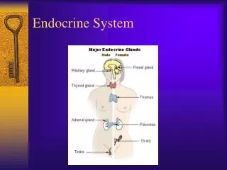
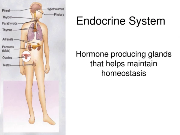
Endocrine System. Background. Hormones. Major Endocrine Organs. Thymus. Control of hormone release. Hormonal activity—half-life, onset and duration. Gonads. Pineal gland. Pituitary (hypophysis). Target cell specificity. Parathyroid glands. Adrenal glands. Pancreas. Chemistry.

Endocrine System
E N D
Presentation Transcript
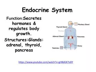
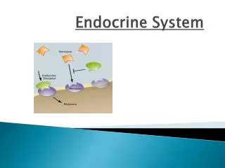
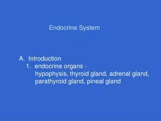
Endocrine System Background Hormones Major Endocrine Organs Thymus Control of hormone release Hormonal activity—half-life, onset and duration Gonads Pineal gland Pituitary (hypophysis) Target cell specificity Parathyroid glands Adrenal glands Pancreas Chemistry Posterior pituitary hormones Thyroid gland Anterior pituitary hormones Mechanism of action—increase or decrease rates of normal cellular activity 1. Endocrine: Ductless, secrete hormones into surrounding tissue fluid, vascular or lymphatic drainage receive hormones, examples of endocrine glands (pituitary, thyroid, parathyroid, adrenal, pineal & thymus) & some organs also have discrete areas of endocrine tissue as well as exocrine tissue (pancreas, gonads & hypothalamus) 2. Types of stimuli: Humoral (glands release hormones in direct response to changing levels of ions or nutrients, e.g., PTH release in response to changes in calcium levels), neural (nerve fibers stimulate hormonal release, e.g., sympathetic activated release of catecholamines from adrenal medulla) & hormonal (release of hormones in response to other hormones, e.g., hypothalamic releasing and inhibiting factors) 3. Overview of second-messenger systems a. Hormone binds plasma membrane receptor b. G-protein signals effector to produce an intracellular message (second messenger) c. Second messenger mediates cellular response to hormone (signaling cascades & protein kinases) 1. Hormonal effects a. Alter plasma membrane permeability b. Alter protein or regulatory molecule synthesis c. Activate or inactivate enzyme d. Induction of secretory activity e. Stimulate mitosis 5. Direct gene activation a. Steroid hormones are lipid soluble (pass through plasma membrane) b. Once inside, hormone binds to intracellular receptor (activated complex is formed) c. Activated complex passes into nucleus and binds to specific DNA sequences d. Association with DNA sequence turns on gene (gene sequence is transcribed) c. Signs of DM: i. Polyuria, ii. Polydipsia & iii. Polyphagia, d. Polyuria: i. Excessive glucose in kidney filtrate acts as a diuretic (i.e., inhibits water reabsortion), ii. Increased urine output causes dehydration & decreased Bd volume, iii. Electrolyte loss with excretion of excess ketones (- charged), e.Polydipsia: dehydration stimulate thirst center in brain, f. Polyphagia: i. Glucose cannot be used because it cannot be absorbed by cells, ii. Results in hunger 8. Gonadotropins: FSH and LH: a. Regulate gonads, b. FSH stimulates gamete production c.LH promotes production of gonadal hormones,d. FSH and LH work in concert to cause follicle to mature (LH causes egg to be extruded from follicle), e. LH stimulates interstitial cells of testes to produce testosterone, f. LH & FSH release is controlled by the hypothalamus (GnRH) & g. Negative feedback inhibition regulates FSH & LH release 2. Connections between posterior pituitary & hypothalamus: a. Posterior is an outgrowth of brain & maintains its neural connections, b. Neurons in supraoptic & paraventricular nuclei of hypothalamus give rise to hypothal-amic-hypophyseal tract (hormones synthesis in secretory cells of hypothalamus: oxytocin & antidiuretic hormone) & c. When neurons fire, hormones are released into capillary bed in posterior pituitary 3. ADH (antidiuretic hormone): a. Inhibits or prevents urine production, b. In response to increases in solute concentration, ADH is released from hypothalamus (hypothalamus has osmoreceptors), c. ADH causes kidney tubules to reabsorb more water, d. At high doses, ADH causes vasoconstriction (Causes increases systolic BP) & e. Diabetes insipidus (tasteless: deficiency in ADH secretion with output of huge amounts of urine & thirst) i. Renin-angiotensin mechanism: JGA release renin in response to BP decrease, initiates cascade forming angiotensin II formation, which stimulates aldosterone release from adrenal cortex,ii. Direct stimulation by plasma sodium and potastium ions, iii. ACTH: at very high levels of ACTH, aldosterone secretion is increased, iv. ANP: Atrial natriuretic peptide: when BP is high, heart release ANP to inhibit renin and aldosterone secretion e. Increases Ca++ absorption by intestine (stimulates conversion of vitamin D into active form), f. Hyperparathyroidism is rare (Ca++ is leached from bones and replaced by connective tissue, elevated blood Ca++ asversely affects NS and contributes to formation of kidney stones as excess Ca++ is deposited in kidney tubules), g.Hypoparathyr-oidism: PTH deficiency following injury or surgical removal (increased NS excitability) 1. Half-life—measure of hormonal persistence in blood stream depends on rate of synthesis and release, speed of removal or degradation 2. Onset of effect is dependent on hormone type (steroid: hours to days) 3. Duration is generally short (e.g 20 minutes) although depends on hormone type 2. Mechanisms that transduce hormonal signal into an intracellular change a. G-protein linked receptor activation of intracellular second messengers (amino acid-based hormones) b. Direct gene activation (steroid hormones) 7. Regulation of insulin: Humoral response to increased circulating glucose 8. Diabetes mellitus (DM): Hyposecretion or hypoactivity of insulin: a. Excessive hyper-glycemia triggers sym. response (activates systems associated with hypoglycemia), b. Lipidemia: i. Fats mobilized to use as cellular food, ii. FA metabolites accumulate as ketone bodies, iii. Bd pH drops (ketoacidosis), 2. Oxytocin: a. Stimulates smooth muscle contraction, b. Muscle response depends on number of oxytocin receptors in uterus and breast (number of receptors increases during pregnancy & afferent impulses as uterus stretches during pregnancy signals release of oxytocin during late stages of pregnancy), c. Hormonal trigger for milk ejection & d. Positive feedback mechanism 5. Growth hormones 1. Same sex hormones as those produced by adrenal cortex 2. Ovaries produce estrogens & progesterone (sexual maturation & menstrual cycle) 3. Testes produce testosterone (sexual maturation & sex drive) 4. Release of gonadal hormones is regulated by gonadotropins 3. Connections between ant. pituitary & hypo-thalamus: a. Anterior lobe is derived from epithelial tissue, b. No direct connection between post. pituitary or hypothalamus, c. Vascular connection (hypophyseal portal veins), d. Releasing & inhibiting hormones secreted by hypothalamus are carried by portal system to anterior pituitary (regulate activity of secretory cells in ant. Pituitary) 1. Typically negative feedback: Hormone secretion is triggered in response to a stimulus & as hormone level increases, target organ is affected & further hormone release is inhibited. 2. Factors affecting target cell activation a. Hormonal levels b. Number of receptors on target cell c. Receptor affinity (can be up or down regulated based on microenvironmental conditions) 3. Effects: Insulin: hypoglycemic hormone & Glucagon: hyperglycemic hormone 4. Glucagon effects:a. Breakdown of glycogen to glucose (glyconeogenesis), b. Synthesis of glucose from lactic acid, fatty acids & amino acids, c. Release of glucose from liver 5. Regulation of glycogen: Humoral response to decreased blood glucose 9. Types of DM: Type I: Insulin dependent DM (IDDM), autoimmume destruction of β cells, juvenile onset, lack insulin activity, long term cardiovascular & neural problems Type II: Non-insulin dependent DM (NIDDM), usually after the age of 40, 90% of DM cases, most patients are overweight, genetic link, insulin is produced in inadequate quantities or with faulty receptors d. Glucocorticoids (type of corticosteroid): i. Influence metabolism and mediate response to stress, ii. Cortisol, cortisone,corticosterone iii.Only cortisol secreted in significant amount iv. Non-stress: CRH, ACTH, cortisol release, negative feedback, v. Stress: Sympathetic NS overrides inhibitory effect of elevated cortisol levels & triggers CRH release, vi. Gluconeo-genesis: Conversion of fats into glucose 1. Two endocrine glands: a. Adrenal medulla (acts as part of the sympathetic NS) & b. Adrenal cortex 2. Response to stressful conditions 3. Adrenal cortex: a. Corticosteroids (steroids, more than two dozen, synthesized from cholesterol), 3. Parathyroid hormone: a. Controls Ca++ balance, b. Released in response to falling blood Ca++ levels, c. PTH stimulates osteo-clast activity (digest bone matrix & release Ca++ ), d. Enhances Ca++ reabsorption by kidney tubules, Classification a. Amino acid-based hormones (most hormones) b. Steroid hormones (gonadal and adrenocortical hormones) 2. Thyroid hormone (TH): a. Two metabolically active iodine-containing hormones: thyroxine (T4) & triiodothyronine (T3), b. Thyroxine (T4) is produced by thyroid gland, c. Triiodo-thyronine (T3) is formed at target tissue (T4 is converted into T3), d. Increases metabolism in most tissues by stimulating glucose oxidation, e. Increases adrenergic receptors in blood vessels, 3. Metabolic disturbances with thyroid gland activity: a. Myxedema: hypothyroid disorder (if from lack of iodine, condition is endemic or colloidal goiter, colloid is made but cannot be iodinated to make functional hormone, TSH secretion increase to stimulateTH production, follicles accumulate more unuseable colloid), 6. Insulin effects: a. Lower blood glucose ( enhance membrane transport of glucose into body cells), b. Alter protein & fat metabolism, c. Inhibits breakdown of glycogen, d. Triggers enzymatic activity (oxidation of glucose for ATP production, synthesis & storage of glycogen, conversion of glucose to fat & its storage 1. General characteristics: a. Connected to hypothalamus (part of brain) by infundibulum (stalklike connection between brain & endo-crine system), b. Two major lobes: Posterior & anterior, c. Posterior lobe + infundibulum = neurohypophysis, d. Anterior lobe (adeno-hyophysis) is comprised of glandular tissue e. Highly vascular f. Regulates tissue growth and development, g. T4 is bound to plasma proteins (TBG: thyroxine-binding globulin) & transported to target tissues(bind target tissue receptors, T3 is bound more readily, h. Regulation: Falling levels trigger TSH release, rising levels of thyroxine inhibits TSH release & conditions in which there is increased energy requirements causes TRH release from hypothalamus • 1. Anterior pituitary is the Master gland • 2. Six hormones as well as a number of other active molecules • 3. Tropic hormones (4/6): Regulate secretory activity of other endocrine glands: • TSH: Thyroid-stimulating hormone • ACTH: Adrenocorticotropic hormone • FSH: Follicle-stimulating hormone • LH: Lutenizing hormone 4. Calcitonin: a. Lowers blood calcium levels, b. Antagonist to the effect of parathyroid hormone: Inhibits calcium release from bones by osteoclast activity & stimulates calcium uptake and incorporation, calcium acts as a humoral signal for calcitonin release b. Mineralocorticoids (type of corticosteroid: Regulate electrolyte concentrations in extra-cellular fluid, aldosterone is most abundant, it reduces excretion of sodium from the body, stimulates reabsortion of sodium in the distal tubule of kidney), c.4 Mechanisms controlling aldosterone secretion g.Gonadocorticoids(Sex hormones): primarily androgens: i. Androstenedione converted to testosterone & dihydrotestosterone, ii. Small amounts of estrogens, iii. Adrenal cortex sex hormones is only fraction of gonadal sources, iv. Possible role in onset of puberty (levels rise during years preceding onset) 1. Structure a. Two lobes connected by isthmus b. Follicles: Follicle cells produce thyroglobin & lumen stores colloid (thyroglobin in association with iodine) c. Thyroid hormone is derived from iodinated thyroglobin d. Parafollicular cells produce calcitonin 1. Two pairs of glands in the posterior aspect of the thyroid gland 2. Chief cells (principal cells) secrete PTH: parathyroid hormone 1. Large in children, decreases with age 2. Hormonal products important for T cell maturation (thymopoietins & thymosins) f. Addison’s disease: hyposecretory disorder of adrenal cortex: i. Weight loss, ii. Reduced plasma glucose & sodium levels, iii. Severe dehydration & hypotension, 1. Mediated by specific protein receptors a. Receptors are localized to cells that are influenced by a given hormone b. Hormones act as molecular triggers 4. Examples of signaling mechanisms a. cAMP b. PIP-Calcium signal mechanism 7. Adrenocorticotropic hormone (ACTH): a. Stimulates adrenal cortex to release cortico-steroid hormones (Glucocorticoids offset effects of stress), b. Its release is controlled by CRH (Corticotropin-releasing hormone: a hypothalamic hormone having a diurnal rhythm) & c. Feedback inhibition: rising glucocorticoids inhibit CRH secretion 1. Contains both exocrine (GI enzymes) & endocrine cells 2. Pancreatic islets (islets of Langerhans) a. Two populations i. Alpha cells—produce glucagons ii. Beta cells—produce insulin • 6. Thyroid-stimulating hormone (TSH) • Stimulates normal growth & activity • of thyroid gland • b. Tropic hormone • c. Controlled by hypothalamus • i. TRH—thyroid releasing hormone • ii. Feedback inhibition • iii. GHIH also inhibits 4. Other hormones (2/6) have neuroendocrine targets: a. PRL: Prolactin b. GH: Growth hormone b. Cretinism: hypothyroidism in infants (TH replacement therapy prevents, cannot reverse effects), c. Graves’ disease: Hyperthyroid pathology, autoimmune disease, abnormal antibodies that mimic TSH, exophthalmus 4. Adrenal medulla (AM): a. Chromaffin cells (Modified postgang. sympathetic neurons that secrete epinephrine & NE), b. Initial response to stress is mediated by sympathetic NS, c. Activation of AM & associated release of EPI & NE prolong sym. response (High BP & HR, mobilization of glucose&shunt blood from GI) e. Cushing’s disease: excess cortisone: i. Characterized by persistent hyperglycemia (steroid diabetes), ii. Loss of muscle & bone protein, iii. Water & salt retention, iv. “moon” face, v. Redistribution of body fat (e.g., buffalo hump), vi. Anti-inflammatory effects mask infection chemical substances secreted by cells into extracellular fluids, that regulate metabolic function of other cells in the body 2. Exocrine: Have ducts & nonhormonal products are directed to membrane surfaces 1. General characteristics a. ADH and oxytocin are comprised of 9 aa (differ only in the identity of 2 residues) b. Released in response to neural signals from hypothalamus f. Effects of growth hormone: Stimulates uptake of amino acids from blood and their incorporation into proteins, stimulates sulfur uptake, mobilizes fats from fat deposits, decreases rate of glucose uptake and metabolism (diabetogenic effect: elevation of blood glucose) There are two types of glands: Endocrine & Exocrine 1. Floor of 3rd ventricle within diencephalons 2. Primary secretory product is melatonin 3. Pineal gland receives indirect inputs from visual system 4. SCN has melatonin receptors h. Abnormalities: *Adolescent hypersecretion: Gigantism *Adult hypersecretion: Acromegaly (tissues still sensitive to GH grow disproportionately) *Adult hyposecretion: Little effect (progeria: occurs when deficit is severe) *Adolescent hyposecretion:Pituitary dwarfism a. Produced by somatotropic cells b. Stimulates most cells to grow & divide c. Major targets are bones and muscles d. Anabolic hormone (promotes metabolism) e. Growth-promoting effects are mediated indirectly (IGFs: Insulin-like growth factors produced by liver and other tissues) 9. Prolaction a. Stimulates milk production b. PRH and PIH (serotonin and dopamine) c. Levels parallel those of estrogen • g. Regulation by hypothalamic hormones (negative feedback): • GHRH: growth hormone releasing hormone (somatocrinin) • GHIH: (growth hormone inhibiting hormone (somatostatin) Home Exit BASIM ZWAIN LECTURE NOTES