ENDOCRINE SYSTEM
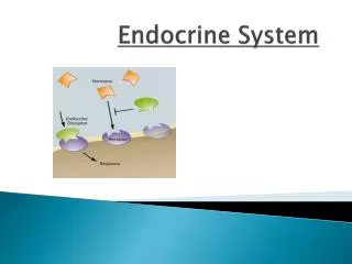
ENDOCRINE SYSTEM. Do not write !!!!!! Everything is available @. http://www.lfhk.cuni.cz/patanat/endocrine. ENDOCRINE SYSTEM. secretion of hormones ( steroids, peptides ) feedback inhibition blood regulation of activity of various organs.


ENDOCRINE SYSTEM
E N D
Presentation Transcript
ENDOCRINE SYSTEM Do not write!!!!!! Everything is available @ http://www.lfhk.cuni.cz/patanat/endocrine
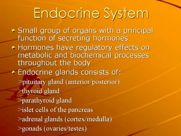
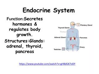
ENDOCRINE SYSTEM secretion of hormones (steroids, peptides) feedback inhibition blood regulation of activity of various organs
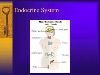
PITUITARY GLAND 0.5 g sella turcica (sphenoidal bone) diaphragma sellae (dura mater) stalk - neural and vascular connection with hypothalamus adeno- neuro- (so called neurohemal organ)
Adenohyphysis ventral lobe embryologically derived from mouth cavity(Rathke's cleft) 5 hormones: ACTH TSH FSH+LH prolactin GH eosinophils (A cells) basophils (B cells) chromophobes
Hyperpituitarism and adenomas of pituitary prolactinoma 30% from A and chromophobes FSH + LH 15% from B ACTH 15% from A and chromophobes GH 5% TSH 1%
Pituitary adenomas majority of adenomas produce only 1 hormone up to 30% adenomas non-functional – only local pressure effect „balloon“ expansion of sella usuration rupture of diaphragm + suprasellar growth pressure to chiasma & n. opticus, impression of brain & paranasal sinuses disturbances of vision, headache X-ray changes carcinoma – very rare
Hormonal syndromes prolactin oligo-, amenorrhea, galaktorrhea, impotence GH gigantism up to 240 cm acromegaly, macroglossy ACTH Cushing's d.: obesity, moon-face, hirsutism, hypertension etc. (see adrenal)
Hypopituitarism loss of at least 75% of parenchyma due to: 1. nonfunctional adenoma (pressure atrophy) 2. ischemic necrosis (Sheehan's sy = post-partum necrosis of enlarged hypophysis by bleeding or haemorr. shock lactation arrest, no restoration of menstrual cycle) 3. empty sella sy – following inflammation, operation, irradiation herniation of arachnoid & CSF into the sella
Hypopituitarism - clinical symptoms •pituitary nanism: decrease of GH substitution •hypogonadism (Fröhlich's sy = dystrophia adiposogenitalis) – accomp. by mental retardation namely in males •hypothyroidism •disorders of adrenal cortex
Posterior lobe syndrome the cause is usually in hypothalamus, very rare decreased ADH diabetes insipidus (polyuria, polydypsia, dehydratation)
Craniopharyngioma = benign tumor from canalis craniopharyngicus remnants (residual structure from Rathke's cleft – source of adenohypophysis) suprasellar expansion often cystic, similar structure as adamantinoma of the jaws
THYROID GLAND regulated by adenohypophysis (TSH) and blood levels of iodine thyroglobulin in follicular colloid transformation to thyroxine (T4) a triiodothyronine (T3) parafollicular C cells calcitonin – facilitates binding of Ca2+ to bones and inhibits bone resorption derived from pharyngeal epithelium – thyroglossal duct persistence thyroglossal duct cyst (median neck cyst) lingual thyroid
Thyroid gland - pathology • more frequent in females (M:F = 1:10!) • namely enlargement - goiter • increased secretion - hyperthyroidism, • thyreotoxicosis • decreased secretion - hypothyroidism • hyperplasia, inflammations, tumors
Hyperthyroidism • diffuse hyperplasia of TG (M. Graves-Basedow) • toxic nodular goiter • toxic adenoma • thyroiditis • pituitary adenoma, hypothalamic disorders
Hyperthyroidism - clinical symptoms • increase of basal metabolism, O2 consumption • restlessness, emotional lability • tremor, sweating, loss of weight, • intolerance of warmth • SOB, increased heart rate and output, palpitations • congestive heart failure due to thyrotoxic • cardiomyopathy (dilated type) • exophtalmus
Hypothyroidism • loss of parenchyma (resection, irradiation, • medication) • Hashimoto's thyroiditis • idiopathic (autoimmune?) hypothyroidism
Hypothyroidism - clinical symptoms IN CHILDHOOD - cretinism endemic iodine deficiency in mountain regions ( addition of iodine to salt) short stature, big tongue, defective teeth, rough facial features IN ADULTHOOD - myxedema accumulation of mucopolysacharides in corium pale thick (doughlike) skin, namely in periorbital areas bradycardia, apathy, intolerance of cold, big lips and tongue enlarged and failing heart with pericardial fluid coronary arteriosclerosis due to hypercholesterolemia
THYROIDITIS •Hashimoto's thyroiditis (= H. goiter) most frequent inflammation, autoimmune immune reaction against TG - up to 20× more frequent in females histology: replacement of parenchyma by lymphoid tissue with formation of lymph. follicles with germinal centers follicular cells eosinophillic, finely granular oncocytes (Hürthle cells), goiter Dx.: clinical symptoms, autoAb., US, (+FNACytology) risk of MALT - lymphoma !
•Focal lymphocytic t. very frequent, in females usually only subclinical manifestation (increase of TSH, normal T3, T4) often incidental morphological finding •Subacute granulomatous t. (De Quervain's) viral etiol.? - fever, palp. tenderness, pain, transitory hyperfunction granulomas with multinucleated giant cells heals spontaneously, not operated •Fibrous goiter (Riedel's) firm idiopathic fibrosis of the gland, merging into surrounding structures, extremely rare
GRAVES - BASEDOW DISEASE • = toxic goiter (pulsans, vibrans et fremens) • most frequent cause of hyperthyroidism (diffuse hyperplasia) • triad: hyperthyroidism • exophthalmia (in 2/3) - edema of retrobulbar • connective tissue • ("malignant" e. – not possible to close eyelids – corneal • ulcers - blindness) • pretibial edema - (in 1/6) - mucin, lymphocytes • up to 7× more frequent in females • autoimmune mechanism (thyroid stimulating Ab., • thyroid growth stimulating Ab.) – against TSH-receptors
GB goiter - histology "too much epithelium, too few colloid" epithelial cells tall, colloid pale, "watery", vacuolated (marginal usurations), stromal lymphoid infiltrates rich vascularization
GOITER this term doesn't say neither anything aboutetiology nor about the character of the process most often of hyperplastic origin first diffuse, later on nodular often accompanied by regressive changes
•Endemic goiter by iodine deficiency decreased synthesis of hormone compensatory increase of TSH enlargement (hyperplasia) of the gland •Sporadic goiter multifactorial, i.e. iodine and goitrogenes in diet: cabbage, cauliflower, turnip, kale females, frequently onset in puberty or pregnancy
Nodular colloidal goiter weight 300g up to 1kg, sometimes retrosternal growth histologically - nodules, sometimes with bleeding and/or calcifications micro- normo- a macrofollicular (majority) - large follicles with colloid (colloidal goiter) eufunctional g., toxic g., hypofunctional g. cytology of cold nodes (diff. from carcinoma) (suspicious goiters and g. with clin. symptoms are operated)
TUMORS 80% of solitary nodules are adenomas benign, mainly solitary, spheric, encapsulated follicular adenoma normofollicular, macrofollicular (colloidal), microfollicular (fetal), trabecular (embryonic) nonfunctional a. (scintigrafic) - cold nodule functional a. – hot nodule (= toxic) oncocytic adenoma large eosinophillic cells
CARCINOMAS • notfrequent, up to 3 × more often in females • post-irradiation- Hiroshima 7% survivors, Tschernobyl, • therapeutic irradiation (lymphomasin • childhood) • from follicular cells • well differentiated - papillary, follicular, oncocytic • poorly differentiated - insular • undifferentiated - anaplastic • from C cells • medullary
•Papillary carcinoma approx. 70% of all carcinomas diagnostic feature is not presence of papillae, but so called „ground glass nuclei" sometimes only minute (mm) - microcarcinoma invasion into capsule, fibrosis psammoma bodies (concentric calcifications) meta to LN, good prognosis - 80% 10y. survival
•Follicular carcinoma about 20% of malignancies difficult diff.dg. vs. adenoma - invasionthrough the capsule and/orvascular invasion! meta to bones, lungs, brain •Anaplastic carcinoma 10% of malignancies, highly agressive histologically – small cell, large cell, spindle cell type death within 2 years •Medullarycarcinoma from C cells (calcitonin!), sometimes familialoccurrence solid foci ofsmall cells, production of amyloid („APUD amyloid")
PARATHYROID GLANDS derived from 3rd and 4thbranchial pouches (together w. thymus) ! ectopy – frequent; supernumerarygland production of parathormone (PTH) : releases Ca2+ from bones increases Ca2+level in serum Hyperparathyroidism primary - cause within the gland secondary - causes outside the gland (e.g. kidneys) hypercalcaemia
Primary hyperparathyroidism • adenoma or hyperplasia • carcinoma (extremely rare) • adenoma mostly solitary, up to 2 cm, encapsulated, • embedded within thyroid, thymus, soft tissue of the neck • histologically chief cells or oxyphillic cells (oncocytes) • treatment = surgery (rarely alcohol injection) • hyperplasia (all 4 glands) – CAVE – MEN! • clinically metastatic calcification, urolithiasis • osteomalacia, ostitis fibrosa cystica (brown tumor of bones)
Secondary hyperparathyroidism compensatory hyperfunction most frequent in: chronic renal insuf. (hyperphosphataemia, hypocalcaemia) hypovitaminosis D (reduced supply of Ca2+ increase of PTH hyperplasia of parathyr. g. - reversible paraneoplastic sy Hypoparathyroidism complication of surgery (thyroidectomy) hypocalcaemia neuromuscular cramps (tetany)
ADRENAL GLANDS 2 organs in 1 cortex vs. medulla different embryogenesis different structure & function
ADRENAL CORTEX spongiocytes producing steroid hormones glucocorticoids, mineralocorticoids, sex steroids hyperfunction, hypofunction, tumors •Hyperfunction (hypercorticism) steroids: glucocorticoids (mainly cortisol) Cushing's sy mineralocorticoids (mainly aldosterone) hyperaldosteronism (Conn's sy) androgens virilism (adrenogenital sy)
Cushing's sy - clinical symptoms • obesity (so called arachnoid type) • moon-face, neck hump, striae • hypertension, muscle weakness • osteoporosis, hirsutism and amenorrhea • impaired metabolism of glucose (steroid diabetes) • psychotic disorders
Cushing's sy - causes • pituitaryadenoma – increase of ACTH • hyperplasia of the cortex • functioning cortical adenoma • paraneoplastic sy - (in 10-15%) – increased ACTH • produced by tumor cells (most often small cell lung cancer) • hyperplasia of the cortex • iatrogenic – Cushing's sy caused by treatment • (glucocorticoids - immunosupression atrophy of • the cortex)
Cushing's sy - morphology •cortical adenoma - high level of cortisol causes hyaline degeneration of B cells in hypophysis Crooke's cells - atrophy of the cortex •pituitary adenoma - hyperplasia of adrenal cortex - diffuse or nodular - bilateral
Hyperaldosteronism mineralocorticoid aldosterone regulation through renin-angiotensin system increased excretion of K+ and retention of Na+ hypokalemia, hypernatremia increased volume of extracellular fluid, blood hypertension muscle weakness (including myocardium) primary aldosteronism in cortical adenoma - Conn's sy
Adrenogenital sy adenoma, hyperplasia or carcinoma of the cortex in young females masculinisation in young males pubertas praecox
Hypofunction primary = insufficiency due to damage of cortex (Addison's disease) secondary = insufficiency due to pituitary lesion ( decrease of ACTH)
Addison's disease decrease of gluco-, mineralocorticoids and androgens damage of at least 90% of parenchyma (bilaterally) in the past majority of cases - TBC epinephritis today usually idiopathic - autoimmune?, amyloidosis, meta, sudden termination of steroid treatment (cortex is atrophic!)
Addison's disease • Morphology • leaf-like adrenals (very thin cortex) • Clinical symptoms • weakness, fatigue, skin and mucosa pigmentation - melanin • hypoglycemia, hypotension • diarrhea, loss of weight • stress may lead to acute crisis with coma (acute • cortical insufficiency) • massive bleeding into cortex (labor trauma, venous • thrombosis, meningoc. sepsis w. DIC – • Waterhause-Friderichsen sy)
Tumors • adenoma – majority non-functional, 1-2 cm • incidental finding in US, CT or at autopsy • histology= zona fasciculata • carcinoma – very rare, usually non-functional • myelolipoma - benign mesenchymal tumor • histology – similar to bone marrow
ADRENAL MEDULLA • chromaffine cells producing epinephrine (adrenalin) • and norepinephrine (noradrenalin) • pathology of medulla: virtually only 2 neoplasms • •Pheochromocytoma • Neuroblastoma
Pheochromocytoma production of adrenalin a noradrenalin 90% from medulla, 10% from sympatic ganglia (paraganglioma) „tumor of 3× 10%": 10% bilateral 10% extraadrenal 10% malignant grams to kg! histology: polygonal cells, EM a immunocytoch. neuroendocrine granules clinically: permanent or paroxysmal hypertension (tachycardia, sweating, headache)
Neuroblastoma highly malignant tumor of children aged 5 - 15 years from adrenal medulla and sympat. ganglia (cervical, thoracic and abdominal) related to retinoblastoma frequent necroses, bleeding and intratumoral calcifications (X-ray!), sometimes production of catecholamins histology: small cells, Homer-Wright rosettes metastases to bones and liver according to degree of differentiation neuroblastoma ganglioneuroblastoma ganglioneuroma
LANGERHANS‘ ISLETS part of APUD system A-cells (glucagon - hyperglycemia) B- cells (insulin) D- cells (somatostatin - „inhibits“ both above mentioned) PP- cells (pancr. polypeptide - activates secretion of various GI enzymes) gastrin – where???
DIABETES MELLITUS !! 5% of population in Czech Republic !! chronic defect of carbohydrates metabolism affects also metabolism of lipids and proteins insufficient production of insulin by B-cells hyperglycemia glycosuria, polyuria (osmotic) causes: idiopathic (genetic) secondary (destruction of L.i. by inflammation, surgery, tumor, hemochromatosis)
Idiopathic DM type I insulin-dependent, juvenile 10% type II insulin-non-dependent, adult 90% genetic disposition obesity (80% DM-II pts. are obese, 60% of obese pts. have disorders of metabolism of carbohydrates) pregnancy, stress, viral infections 7th most frequent cause of death – increasing tendency!
Pathogenesis of DM insulin regulates: utilisation of glucosis in cells synthesis of glycogen (liver and muscles) synthesis of triglycerides from glucose synthesis of proteins lack of insulin hyperglycemia glycosuria + ketosis + acidosis intoxication by ketones diabetic coma DM I - insulin is missing B cells destroyed by autoimmunity, viral infection, ??? survival - exogenous insulin (insulin-dependent DM) DM II – mildly impaired secretion + resistance of peripheral cells to insulin
Morphology • •Pancreas– changes of L. islets • often none • sometimes reduction of size and/or number • sometimes increase of size and/or number • - babies of diabetic mothers • less frequently APUD amyloid in L.i. • degranulation of B cells (EM) • lymphocytic infiltration of L.i. („insulitis")