Download

1 / 34
380 likes | 724 Vues
A brain abscess is a localized infection in the brain, resulting from microorganisms entering the cerebral tissues due to trauma, contiguous infections, or hematogenous spread from distant sites. Common sources include frontal sinus infections, middle ear infections, and hematogenous dissemination from conditions such as pulmonary infections and osteomyelitis. Symptoms may include fever, neurological deficits, and raised intracranial pressure. Diagnosis involves imaging techniques like CT and MRI. Management typically includes antibiotics and may require surgical intervention, especially for larger abscesses.

E N D
CNS INFECTIONS – BRAIN ABSCESS Prof.Arjun Shetty Dept Of Neurosurgery Yenepoya Medical College
Brain abscess • Occurs when micro organisms are introduced into the cerebral tissues commonly as a result of • 1.trauma • 2.contiguous infection • 3.hematogenous dissemination of infection
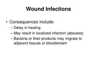
Frontal sinus infection • Retrograde thrombophlebitis of diploic vein • Direct extension secondary to osteomyelitis • Abscess in frontal lobe base
Middle ear infection • Direct spread through tegmen tympani • Trans-labyrinthine spread through round/oval window • Direct spread of mastoid sinus infection • Abscess in temporal lobe • Abscess secondary to mastoiditis can occur in the cerebellum
Abscesses due to sinusitis or otitis tend to be single and superficial
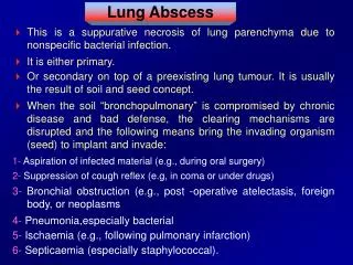
Hematogenous spread of infection • Skin pustules • Pulmonary infection • Osteomyelitis • Dental abscess • Diverticulitis • Infective bacterial endocarditis
Hematogenous abscesses occur usually in M.C.A territory • These abscesses occur in the cortico medullary junction – slow blood flow • Can be multiple • Blood brain barrier usually prevents abscess formation secondary to transient bacteremia
Brain abscess can be associated with cyanotic heart disease especially right to left shunt
Right to left shunt • Pulmonary capillary filtration is bypassed • Hypoxia leads to polycythemia and increased blood viscosity which causes micro infarcts – nidus for infection
Abscess secondary to cyanotic heart disease have usually single unlike those associated with bacterial endocarditis • Fallot’s tetrology – 50% • Transposition of great vessels • Tricuspid atresia • ASD , VSD
microbiology • In the pre-antibiotic era - staph aureus • Anaerobes are now becoming common • Bacteroids • Anaerobic streptococci • Peptococcus • actinomycosis
aerobes • Staph aureus • Streptococcus enterobactericiae • Haemophilus • 1/3 cases have mixed infection especially secondary to otogenic infection • Neonates – proteus and citrobacter
Immunocompromised patients • Fungal infections – candida,aspergillus,cryptococcus neoformans • Toxoplasmosis – commonest non viral infection in HIV patients
pathology • Stage of early cerebritis : • 1-3 days • necrotic center • local inflammation around the vessels
Stage of late cerebritis : • 4-9 days • Increased necrotic center • Zone of inflammatory cells and macrophages • Fibroblasts lay down reticulin
Stage of early capsule formation : • Day 10-13 • Reticulin network forms capsule
Stage of late capsule formation : • >14 days • Necrotic center • Inflammatory zone and fibroblasts • Capsule • Neo-vasculature surrounding capsule • Zone of edema and reactive gliosis
Clinical features • Male predominance • Symptoms of raised ICP • Fever – tends to be low grade – 50% may be associated with meningeal signs • Focal neurological deficits • Seizures ( 30-50%)
investigations • Laboratory : • 1.WBC count – normal or mildly elevated • 2.ESR – raised, • polycythemia decreases it hence it is unreliable in cyanotic heart disease • 3.lumbar puncture – avoid in SOL with increased ICP, significant only in meningitis
Radiological : • 1.X-ray – PNS , mastoid evaluation for sinusitis, evidence of osteomyelitis , trauma
CT Early cerebritis – hypo dense area with minimal contrast enhancement Late cerebritis – hypo dense area with ring enhancement which diffuses medially Early capsule – well defined ring enhancement with hypo dense center Late capsule – hyper dense capsule demonstrated with non contrast films
MRI • T1 – central hypo intensity , capsules are iso to hyper intense , edema is hypo intense • T2 – necrotic area is iso to hyper intense,capsule is hypo intense,edema is hyper intense
Differential diagnosis • Glioma , metastasis , resolving hematoma , radiation necrosis • Ir 111 labelled radionucleotide scan – useful to differentiate abscess from malignant lesion (sensitivity 100% , specificity 96%)
antibiotics • Antibiotics alone – empirical • Not effective in abscess above 2.5cm
Initial treatment • Penicillins , metronidazole ,3rd gen.cephalosporins – covers anaerobes,streptococci,gram negative aerobes • Later antibiotics to be continued as per c/s
Duration of antibiotics – usually 6 weeks to 3 months • If follow up scan shows residual contrast enhancement, follow up CT recommended for every 3 months.antibiotics are reinstated if enhancement increases • Residual tumor enhancement in itself does not indicate need to continue antibiotics • Stoppage of steroids may be associated with an increase in contrast enhancement – this does not indicate re-growth of abscess
Corticosteroids : • 1.decreases edema • 2.decreases mass effect • 3.used only in cases where mass effect is significant problem
Surgery – excision vs aspiration • Excision : • Traumatic abscess with retained fragments • Fungal abscess • Multiple loculated abscesses
Aspiration : • In cerebritis stage • Abscess in deep seated areas • Cyanotic heart disease
sequelae • Epilepsy – 20 to 30% , increased association with excision than aspiration • Hemiparesis & cognitive deficits – increased in excision • Children fare poorly
Abscess in immunocompromised patients: • Toxoplasma gondii – pyrimethamine +sulfamethoxazole+folinic acid • Cryptococcal infection – amphotericin B +s.tancytosine • Pseudomonas – amikacin or tobramicin + cephalosporin • Multiple abscesses without bacteriological diagnosis – may need upto 3 months of iv antibiotics