Download
1 / 85
850 likes | 911 Vues
Explore the cellular basis of reproduction and inheritance, from sea star development to cell division in prokaryotes and eukaryotes. Learn the intricate processes of mitosis and cytokinesis, bridging the connection between cell division and reproduction.

E N D
How to Make a Sea Star — With and Without Sex • The life cycle of a multicellular organism includes • development • reproduction • This sea star embryo (morula) shows one stage in the development of a fertilized egg • The cluster of cells will continue to divide as development proceeds
This sea star is regenerating a lost arm • Regeneration results from repeated cell divisions • Some organisms can also reproduce asexually
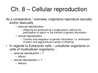
CONNECTIONS BETWEEN CELL DIVISION AND REPRODUCTION • Cell division is at the heart of the reproduction of cells and organisms • Organisms can reproduce sexually or asexually
8.1 Like begets like, more or less • Some organisms make exact copies of themselves, asexual reproduction • One parent = identical copies (see like begets like) Figure 8.1A
Other organisms make similar copies of themselves in a more complex process, sexual reproduction • Two parents = genetic diversity enhances variation • Siblings can have identical and non-identical genetic variants Figure 8.1B
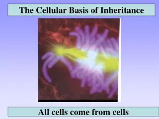
8.2 Cells arise only from preexisting cells • All cells come from cells – “Virchow” • Cellular reproduction is called cell division • Cell division allows an embryo to develop into an adult • It also ensures the continuity of life from one generation to the next
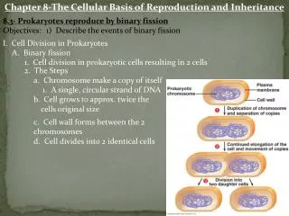
8.3 Prokaryotes reproduce by binary fission • Review Prokaryotic vs. Eukaryotes • Prokaryotic cells divide asexually • These cells possess a single chromosome, containing genes (remember from ch. 4 – no nucleus contained within own nuclear envelop/membrane) • The chromosome is replicated • The cell then divides into two cells, a process called binary fission Prokaryotic chromosomes Figure 8.3B
Plasmamembrane Prokaryoticchromosome Cell wall • Binary fission of a prokaryotic cell Duplication of chromosomeand separation of copies Continued growth of the cell and movement of copies Division intotwo cells Figure 8.3A
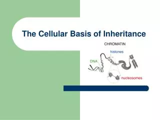
THE EUKARYOTIC CELL CYCLE AND MITOSIS 8.4 The large, complex chromosomes of eukaryotes duplicate with each cell division • A eukaryotic cell has many more genes than a prokaryotic cell • The genes are grouped into multiple chromosomes, found in the nucleus • The chromosomes of this plant cell are stained dark purple Figure 8.4A
Individual chromosomes are only visibleduring cell division • They are packaged as chromatin • Chromosomes contain a very long DNA molecule with thousands of genes
Sister chromatids • Before a cell starts dividing, the chromosomes are duplicated • This process produces sister chromatids • They are linked at centromere (DNA mass of proteins) Centromere Figure 8.4B
Chromosomeduplication • Two daughter cells are produced • Each has a complete and identical set of chromosomes • When the cell divides, the sister chromatids separate Sister chromatids Centromere Chromosomedistributiontodaughtercells Figure 8.4C
8.5 The cell cycle multiplies cells • The cell cycle consists of two major phases (covered in detail in computer lab): • Interphase, where chromosomes duplicate and cell parts are made • G1, S, G2, G0 • Go not listed • The mitotic phase, when cell division occurs Figure 8.5
8.6 Cell division is a continuum of dynamic changes • Eukaryotic cell division consists of two stages: • Mitosis – nuclear/chromosomal division • Cytokinesis – cytoplasmic division (including, but never shown, division of all organelles) • Why?
After the chromosomes coil up, a mitotic spindle moves them to the middle of the cell • Warning: Need to read yellow boxes for flashcard information!!! • In mitosis, the duplicated chromosomes are distributed into two daughter nuclei
INTERPHASE PROPHASE Centrosomes(with centriole pairs) Early mitoticspindle Centrosome Fragmentsof nuclearenvelope Kinetochore Chromatin Centrosome Spindlemicrotubules Nucleolus Nuclearenvelope Plasmamembrane Chromosome,consisting of twosister chromatids Figure 8.6
The process of cytokinesis divides the cell into two genetically identical cells • The sister chromatids then separate and move to opposite poles of the cell
METAPHASE ANAPHASE TELOPHASE AND CYTOKINESIS Cleavagefurrow Nucleolusforming Metaphaseplate Nuclearenvelopeforming Spindle Daughterchromosomes Figure 8.6 (continued)
PMAT • “P” for preparation (prophase)”j • “M” for middle (metaphase) • “A” for away/apart { chromosomes not at poles} (anaphase) • “T” for two, end up with two cells {chromosomes at poles} (telophase) • Telophase concurrently with cytokinesis
8.7 Cytokinesis differs for plant and animal cells • In animals, cytokinesis occurs by cleavage • This process pinches the cell apart Cleavagefurrow Cleavagefurrow Contracting ring ofmicrofilaments Figure 8.7A Daughter cells
Cell plateforming Wall ofparent cell Daughternucleus • In plants, a membranous cell plate splits the cell in two • Notice integrity of cell wall Cell wall New cell wall Vesicles containingcell wall material Cell plate Daughtercells Figure 8.7B
8.8 Anchorage, cell density, and chemical growth factors affect cell division • Most animal cells divide only when stimulated, and others not at all • In laboratory cultures, most normal cells divide only when attached to a surface • They are anchorage dependent
This is called density-dependent inhibition • Cells continue dividing until they touch one another Cells anchor to dish surface and divide. When cells have formed a complete single layer, they stop dividing (density-dependent inhibition). If some cells are scraped away, the remaining cells divide to fill the dish with a single layer and then stop (density-dependent inhibition). Figure 8.8A
Growth factors are proteins secreted by cells that stimulate other cells to divide (Again, think back to computer lab with cell cycle game. After forming a single layer, cells have stopped dividing. Providing an additional supply of growth factors stimulates further cell division. Figure 8.8B
8.9 Growth factors signal the cell cycle control system • Proteins within the cell control the cell cycle • Signals affecting critical checkpoints determine whether the cell will go through a complete cycle and divide G1 checkpoint Controlsystem M checkpoint Figure 8.9A G2 checkpoint
The binding of growth factors to specific receptors on the plasma membrane is usually necessary for cell division Growth factor Plasma membrane Relayproteins G1 checkpoint Receptor protein Signal transduction pathway Cell cyclecontrolsystem Figure 8.8B
Cells - again review cell computer lab. • G1 • G0 – i.e. mature nerve and muscle cells • S – What happens if this phase doesn’t occur? Think chromosome number • G2 • Mitosis/Cytokinesis
8.10 Connection: Growing out of control, cancer cells produce malignant tumors • Cancer cells have abnormal cell cycles • They divide excessively and can form abnormal masses called tumors • Not following density-dependency • Radiation and chemotherapy are effective as cancer treatments because they interfere with cell division
Malignant tumors can invade other tissues and may kill the organism. Benign are localized, not spreading. Lymphvessels Tumor Glandulartissue Metastasis 1 A tumor grows from a single cancer cell. 2 Cancer cells invade neighboring tissue. 3 Cancer cells spread through lymph and blood vessels to other parts of the body. Figure 8.10
It’s Mitosis Time, C’mon! Sing along byAnnette M. Parrott www.africangreyparrott.com
It’s Mitosis Time! C’mon! It’s Mitosis Time! C’mon!
The Cell Cycle’s goin’ on right now In all your cells and you’re gonna find out how...
After Interphase, when the cell has grown again synthesis is done and mitosis begins
It’s Mitosis! Reproduction and it’s time to divide
It’s Mitosis! only in (2n) diploid cells 2 clones side by side
Prophase: nucleus disappears centrioles migrate chromosomes appear
Metaphase: Anaphase: Telophase:
they’re two! = Yahoo!
It’s mitosis time! C’mon! It’s mitosis time! C’mon!
Now we’ll see how much you’ve learned so raise your hand and wait your turn...
Prophase? Metaphase? Anaphase? Telophase? 1 3 2 4 5 6
Prophase? Metaphase? Anaphase? Telophase? 1 2 3 4 5 6 7
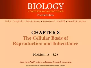
8.11 Review of the functions of mitosis: Growth, cell replacement, and asexual reproduction • When the cell cycle operates normally, mitotic cell division functions in: • Growth (seen here in an onion root) Figure 8.11A
Deadcells • Cell replacement (seen here in skin) Epidermis, the outer layer of the skin Dividingcells Dermis Figure 8.11B
Asexual reproduction (seen here in a hydra) Figure 8.11C