Review of the Microscope
480 likes | 751 Vues
Review of the Microscope. Ch. 1 Biology. Microscopy. Things are not all the same size, are they? Figure shows the resolution of the naked eye, optical (light) microscopy and electron microscopy. History of Microscopy.

Review of the Microscope
E N D
Presentation Transcript
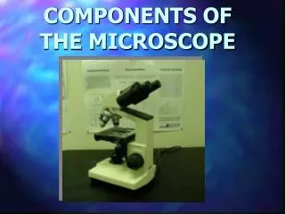
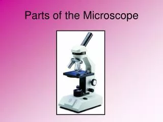
Review of the Microscope Ch. 1 Biology
Microscopy Things are not all the same size, are they? Figure shows the resolution of the naked eye, optical (light) microscopy and electron microscopy.
History of Microscopy • Founding Fathers Of Microscopy:Hans and Zacharias Janssen, ~1590, Dutch Eyeglass Makers, Inventors Credit for the first microscope is usually given to Zacharias Janssen, pictured at the left, in Middleburg, Holland, around the year 1595. Since Zacharias was very young at that time, it's possible that his father Hans made the first one, but young Zach took over the production. • The first compound microscopes produced by the Janssen's was simply a tube with lenses at each end. The magnification of these early scopes ranged from 3X to 9X, depending on the size of the diaphragm openings.
History of Microscopy • Anton van Leeuwenhoek, 1632-1723, Wine Assayer, Surveyor, Cloth Merchant, Minor Public Official, and Inventor Leeuwenhoek was a man with many talents, his most important attributes were creativity, power of observation, and ingenuity. Leeuwenhoek was a common man without any fortune or formal education, so he had to work for a living. Leeuwenhoek made simple (one lens) microscopes. He was not the first person to build a microscope, but the microscopes that he did build were the best ones for that time period. Leeuwenhoek was the first person to describe bacteria (from teeth scrapings), protozoans (from pond water), helped to prove the theory of blood circulation. He gained much of his inspiration form reading Hooke's Micrographia.
Microscopy: The Instruments • A simple microscope has only one lens. Figure 1.2b
Discovery of Microorganisms • Antony van Leeuwenhoek (1632-1723) • first person to observe and describe micro-organisms accurately Figure 1.1b
History of Microscopy • Robert Hooke, 1635-1703, English Chemist, Mathematician, Physicist, and Inventor Hooke's remarkable engineering abilities enabled him to invent and improve many mechanical devices, including timepieces (for which he invented the spiral spring), the quadrant, and the Gregorian telescope. Perhaps even more intriguing than his actual inventions are the devices he designed but never built: he anticipated the invention of the steam engine, and as early as 1684 he described a working telegraph system. • Hooke balanced his inventions with more pure research. Hooke improved on early compound microscopes around 1660. In Micrographia (1665), he coined the word cell to describe the features of plant tissue (cork from the bark of an oak tree) he was able to discover under the microscope. He put his extensive mathematical knowledge in formulating the theory of planetary movement, which provided a basis for Sir Isaac Newton's theories of gravitation. In 1667 he discovered the role of oxygenation in the respiratory system.
The Light Microscope • many types • bright-field microscope • dark-field microscope • phase-contrast microscope • fluorescence microscopes • are compound microscopes • image formed by action of 2 lenses
Microscopy: The Instruments • In a compound microscope the image from the objective lens is magnified again by the ocular lens. • Total magnification =objective lens ocular lens Figure 3.1b
working distance • distance between the front surface of lens and surface of cover glass or specimen
Microscope Resolution • ability of a lens to separate or distinguish small objects that are close together • wavelength of light used is major factor in resolution shorter wavelength greater resolution
The Bright-Field Microscope • produces a dark image against a brighter background • has several objective lenses • parfocal microscopes remain in focus when objectives are changed • total magnification • product of the magnifications of the ocular lens and the objective lens
The Dark-Field Microscope • produces a bright image of the object against a dark background • used to observe living, unstained preparations
The Phase-Contrast Microscope • enhances the contrast between intracellular structures having slight differences in refractive index • excellent way to observe living cells The intact chloroplasts and broken chloroplasts can be differentiated by phase contrast microscopy:
The Differential Interference Contrast Microscope • creates image by detecting differences in refractive indices and thickness of different parts of specimen • excellent way to observe living cells
Differential Interference Contrast Microscopy • Accentuates diffraction of the light that passes through a specimen; uses two beams of light. Figure 3.5
Fluorescence Microscopy • Uses UV light. • Fluorescent substances absorb UV light and emit visible light. • Cells may be stained with fluorescent dyes (fluorochromes). Figure 3.6b
Fluorescence Microscopy Is Widely Used to Visualize Specific Cellular Structures and to Localize Proteins
A Multiprobe Fluorescent Micrograph of a Mitotic Cell Microtubules: green DNA: blue centromeres; red
Newer Techniques in Microscopy • confocal microscopy and scanning probe microscopy • have extremely high resolution • can be used to observe individual atoms Figure 2.20
Confocal Microscopy • confocal scanning laser microscope • laser beam used to illuminate spots on specimen • computer compiles images created from each point to generate a 3-dimensional image
Scanning Probe Microscopy • scanning tunneling microscope • steady current (tunneling current) maintained between microscope probe and specimen • up and down movement of probe as it maintains current is detected and used to create image of surface of specimen
Scanning Probe Microscopy • atomic force microscope • sharp probe moves over surface of specimen at constant distance • up and down movement of probe as it maintains constant distance is detected and used to create image
Electron Microscopy • beams of electrons are used to produce images • wavelength of electron beam is much shorter than light, resulting in much higher resolution Figure 2.20
The Scanning Electron Microscope • uses electrons reflected from the surface of a specimen to create image • produces a 3-dimensional image of specimen’s surface features
Scanning Electron Microscopy (SEM) Visualizes Surface Features Specimens are coated with metals to deflect electrons from a beam scanned across the sample.
SEM of Stereocilia Projecting from a Cochlear (inner ear) Hair Cell
The Transmission Electron Microscope • electrons scatter when they pass through thin sections of a specimen • transmitted electrons (those that do not scatter) are used to produce image • denser regions in specimen, scatter more electrons and appear darker
In Transmission Electron Microscopy, Magnetic Lenses Replace Glass As a Means of Focusing Electromagnetic Waves The shorter wavelength of the accelerated electrons permits much higher resolution.
TEMs Must be Carefully Interpreted Imagine all the misleading interpretations of this branched mitochondrion possible from viewing a single section.
Differential-interference-contrast (Nomarski) Dark field Comparison of Microscopic Techniques for Visualizing a Living, Unstained Cell
Bright field Phase-contrast Comparison of Microscopic Techniques for Visualizing a Living, Unstained Cell
Micro$cope$ • Costs • $150 - $10,000(Compound) • $100-$1500 (Dissection or Stereoscope) • $20,000-100,000 (ConfocalMicroscope) • more than $50.000 (Scanning Electron Microscope(SEM)Transmission Electron Microscope (TEMMicroscope)