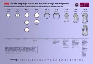
TS 1 (0-2.5)
Theiler Stage dpc. 1 1. 2 1. 3 2. 4 3. 5 4. 6 4.5. 7 5. 8 6. 9 6.5. 10 7. 11 7.5. 12 8. 13 8.5. 14 9. 15 9.5. 16 10. 17 10.5. 18 11. 19 11.5. 20 12. 21 13. 22 14. 23 15. 24 16. 25 17. 26 18. 27 19.

TS 1 (0-2.5)
E N D
Presentation Transcript
Theiler Stage dpc 1 1 2 1 3 2 4 3 5 4 6 4.5 7 5 8 6 9 6.5 10 7 11 7.5 12 8 13 8.5 14 9 15 9.5 16 10 17 10.5 18 11 19 11.5 20 12 21 13 22 14 23 15 24 16 25 17 26 18 27 19 Theiler Staging Criteria for Mouse Embryo Development For full descriptions of Theiler staging and mouse anatomy visit http://genex.hgu.mrc.ac.uk/Atlas/intro.html TS 1 (0-2.5) TS 2 (1-2.5) TS 3 (1-3.5) TS 4 (2-4) TS 5 (3-5.5) TS 6 (4-5.5) TS 7 (4.5-6) TS 8 (5-6.5) TS 9 (6.25-7.25) a b One-cell egg Dividing egg 2-4 cells Morula 4-16 cells Blastocyst, inner cell mass apparent 16-40 cells Blastocyst (zona-free) Attachment of blastocyst, primary endoderm covers blastocoelic surface of inner cell mass Implantation and formation of egg cylinder. Ectoplacental cone appears, enlarged epiblast, primary endoderm lines mural trophectoderm Differentiation of egg cylinder. Implantation sites 2x3mm. Ectoplacental cone region invaded by maternal blood, Reichertís membrane and proamniotic cavity form First sign of radial asymmetry. a Pre-streak(PS), advanced endometrial reaction, ectoplacental cone invaded by blood, extraembryonic ectoderm, embryonic axis visible. b Early streak (ES), gastrulation starts, first evidence of mesoderm. Figures represent development of F1 hybrid (C57BL X CBA) mice. Descriptions of embryos at TS9 – 11 based on Downs & Davies, 1993, and apply to outbred mice of the PO strain. Numbers in parentheses under Theiler stages represent approximate dpc range. Relative sizes of illustrations not to scale. The cartoons are intended as a illustrative guide only.
Theiler Stage dpc 1 1 2 1 3 2 4 3 5 4 6 4.5 7 5 8 6 9 6.5 10 7 11 7.5 12 8 13 8.5 14 9 15 9.5 16 10 17 10.5 18 11 19 11.5 20 12 21 13 22 14 23 15 24 16 25 17 26 18 27 19 Theiler Staging Criteria for Mouse Embryo Development For full descriptions of Theiler staging and mouse anatomy visit http://genex.hgu.mrc.ac.uk/Atlas/intro.html TS 10 (6.5-7.75) TS 11 (7.25-8) TS 12 (7.5-8.75) TS 13 (8-9.25) TS 14 (8.5-9.75) TS 15 (9-10.25) TS 16 (9.5-10.75) TS 17 (10-11.25) TS 18 (10.5-11.25) a a b b c c Turning of the embryo, 1st branchial arch has maxillary and mandibular components, 2nd arch present 8-12 somites. Absent 3rd branchial arch Formation & closure of ant. neuropore, otic pit indented but not closed, 3rd branchial arch visible 13-20 somites. Absent forelimb bud Formation of post. neuropore, forelimb bud, forebrain vesicle subdivides 21-29 somites. Absent hindlimb bud, Rathke's pouch Posterior neuropore closes, Formation of hindlimb & tail buds, lens plate, Rathke's pouch; the indented nasal processes start to form 30-34 somites. Absent thin & long tail Deep lens indentation, adv. devel. of brain tube, tail elongates and thins, umbilical hernia starts to form 35-39 somites. Absent nasal pits Closure of lens vesicle, nasal pits, cervical somites no longer visible 40-44 somites. Absent auditory hillocks, anterior footplate d a Mid streak (MS), amniotic fold starts to form. b Late streak, no bud (LSOB), exocoelom. c Late streak, early bud (LSEB), allantoic bud first appears, node, amnion closing First appearance of somites. 1-4 somites, allantois extends, 1st branchial arch, heart starts to form, foregut pocket visible, preotic sulcus (at 2-3 somite stage). 5-7 somites, allantois contacts chorion at the end of TS12 Absent 2nd arch, >7 somites a Neural plate (NP), head process developing, amnion complete b Late neural plate (LNP), elongated allantoic bud c Early head fold (EHF) d Late head fold (LHF), foregut invagination
Theiler Stage dpc 1 1 2 1 3 2 4 3 5 4 6 4.5 7 5 8 6 9 6.5 10 7 11 7.5 12 8 13 8.5 14 9 15 9.5 16 10 17 10.5 18 11 19 11.5 20 12 21 13 22 14 23 15 24 16 25 17 26 18 27 19 Theiler Staging Criteria for Mouse Embryo Development For full descriptions of Theiler staging and mouse anatomy visit http://genex.hgu.mrc.ac.uk/Atlas/intro.html TS 19 (11-12.25) TS 20 (11.5-13) TS 21 (12.5-14) TS 22 (13.5-15) TS 23 TS 24 TS 25 TS 26 TS 27 Lens vesicle completely separated from the surface epithelium. Anterior, but no posterior, footplate. Auditory hillocks first visible 45-47 somites. Absent retinal pigmentation and sign of fingers Earliest sign of fingers (splayed-out), posterior footplate apparent, retina pigmentation apparent, tongue well-defined, brain vesicles clear 48-51 somites. Absent 5 rows of whiskers, indented anterior footplate Anterior footplate indented, elbow and wrist identifiable, 5 rows of whiskers, umbilical hernia now clearly apparent 52-55 somites. Absent hair follicles, fingers separate distally Fingers separate distally, only indentations between digits of the posterior footplate, long bones of limbs present, hair follicles in pectoral, pelvic and trunk region 56-~60 somites. Absent open eyelids, hair follicles in cephalic region Fingers & toes separate, hair follicles also in cephalic region but not at periphery of vibrissae, eyelids open. Absent nail primordia, fingers 2-5 parallel Repositioning of umbilical hernia, eyelids closing, fingers 2-5 are parallel, nail primordia visible on toes. Absent wrinkled skin, fingers & toes joined together Skin is wrinkled, eyelids are closed,umbilical hernia is gone. Absent ear extending over auditory meatus, long whiskers Long whiskers, eyes barely visible through closed eyelids, ear covers auditory meatus Newborn mouse