Download
1 / 100
1k likes | 1.18k Vues
Biology 3201 Unit 1 Maintaining Dynamic Equilibrium II. Chapter 12 The Nervous System Ms. K. Morris 2010-2011. 12.1 - The Structure of the Nervous System The human nervous system is very important in helping to maintain the homeostasis (balance) of the human body.

E N D
Biology 3201Unit 1Maintaining Dynamic Equilibrium II Chapter 12 The Nervous System Ms. K. Morris 2010-2011
12.1 - The Structure of the Nervous System • The human nervous system is very important in helping to maintain the homeostasis (balance) of the human body. • The human nervous system is a high speed communication system to and from the entire body.
A series of sensory receptors work with the nervous system to provide information about changes in both the internal and external environments. • The human nervous system is a complex of interconnected systems in which larger systems are comprised of smaller subsystems each of which have specific structures with specific functions.
There are two major parts of the human nervous system : (A) Central Nervous System ( CNS ) (B) Peripheral Nervous System ( PNS ) • The CNS is made up of the brain and spinal cord. • The PNS is made up of all the nerves that lead into and out of the CNS. See Fig. 12.2 , P. 392.
The Central Nervous System • The CNS, brain and spinal cord, receives sensory information and initiates ( begins ) motor control. • This system is extremely important and therefore must be well protected. Protection is provided in a variety of ways: • Bone provides protection in the form of a skull around the brain and vertebrae around the spinal cord. • Protective membranes called meninges surround the brain and spinal cord. • Cerebrospinal fluid fills the spaces between the meninges membranes to create a cushion to further protect the brain and spinal cord.
The spinal cord extends through the vertebrae, up through the bottom of the skull, and into the base of the brain. • The spinal cord allows the brain to communicate with the PNS. • A cross section of the spinal cord shows that it contains a central canal which is filled with cerebrospinal fluid, and two tissues called grey matter and white matter. { See Fig. 12.4, P. 393 }
The grey matter is made of neural tissue which contains three types of nerve cells or neurons: 1. Sensory neurons 2. Motor neurons 3. Interneurons • Grey matter is located in the center of the spinal cord in the shape of the letter H. • The white matter of the spinal cord surrounds the grey matter. It contains bundles of interneurons called tracts.
The Peripheral Nervous System • As stated before, the PNS is made up of nerves. • The PNS is made up of two subsystems: 1. Autonomic Nervous System 2. Somatic Nervous System • The autonomic nervous system is not consciously controlled and is often called an involuntary system. It is made up of two subsystems: 1. Sympathetic Nervous System 2. Parasympathetic Nervous System
The sympathetic and parasympathetic systems control a number of organs within the body. • The sympathetic nervous system sets off what is known as a “fight - or - flight” reaction. This prepares the body to deal with an immediate threat. • Stimulation of the sympathetic nervous system causes a number of things to occur in the body: - Heart rate increases - Breathing rate increases - Blood sugar is released from the liver to provide energy which will be needed to deal with the threat.
The parasympathetic nervous system has an opposite effect to that of the sympathetic nervous system. When a threat has passed, the body needs to return to its normal state of rest. • The parasympathetic system does this by reversing the effects above: - Heart rate decreases (slows down) - Breathing rate decreases (slows down) - A message is sent to the liver to stop releasing blood sugar since less energy is needed by the body
The somatic nervous system is made up of sensory nerves and motor nerves. • Sensory nervescarry impulses (messages) from the body’s sense organs to the central nervous system. • Motor nervescarry messages from the central nervous system to the muscles. • To some degree, the somatic nervous system is under conscious control. • Another function of the somatic nervous system is a reaction called a reflex.
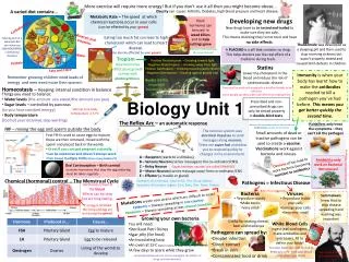
Neurons & Reflex Responses • The neuron or nerve cell is the structural and functional unit of the nervous system. • Both the CNS and the PNS are made up of neurons. • 90% of the body’s neurons are found in the CNS. • Neurons held together by connective tissue are called nerves. • The nerve pathway which leads from a stimulus to a reflex action is called a reflex arc.
A typical nerve cell or neuron consists of three parts: 1. The Cell Body 2. Dendrites 3. Axon (See Fig. 12.6, P. 395) • The cell body is the largest part of a neuron. It has a centrally located nucleus which contains a nucleolus. It also contains cytoplasm as well as organelles such as mitochondria, lysosomes, golgi bodies, and endoplasmic reticulum.
The dendrites receive signals from other neurons. The number of dendrites which a neuron has can range from 1 to 1000’s depending on the function of the neuron. • The axon is a long cylindrical extension of the cell body. It can range from 1mm to 1m in length. When a neuron receives a stimulus the axon transmits impulses along the length of the neuron. At the end of the axon there are specialized structures which release chemicals that stimulate other neurons or muscle cells.
There are three types of neurons: 1. Sensory neuron 2. Motor neuron 3. Interneuron • A sensory neuron carries information from a sensory receptor to the CNS. • A motor neuron carries information from the CNS to an effector such as a muscle or gland. • An interneuron receives information from sensory neurons and sends it to motor neurons. - See Fig. 12.7, P. 396
Complete Core Lab #1 • The Nervous System & Reflex Responses (p.396-397)
The Brain & Homeostasis Today, scientists have a lot of information about what happens in the different parts of the brain; however they are still trying to understand how the brain functions. • We know that the brain coordinates homeostasis inside the human body. It does this by processing information which it receives from the senses. • The brain makes up only 2 percent of the body’s weight, but can contain up to 15 percent of the body’s blood supply, and uses 20 percent of the body’s oxygen and glucose supply. • The brain is made up of 100 billion neurons.
Early knowledge of how the brain functions came from studying the brains of people who have some brain disease or brain injury. Today, innovations in technology have resulted in many ways of probing the structure and function of the brain. These include: 1)The electroencephalograph ( EEG ) which was invented in 1924 by Dr. Hans Borger. This device measures the electrical activity of the brain and produces a printout ( See Fig. 12.8, P.398 ). This device allows doctors to diagnose disorders such as epilepsy, locate brain tumors, and diagnose sleep disorders.
2) Another method is direct electrical stimulation of the brain during surgery. This has been used to map the functions of the various areas of the brain. In the 1950s, Dr. Wilder Penfield, a Canadian neurosurgeon was a pioneer in this field of brain mapping
3) Advances in scanning technology allow researchers to observe changes in activity in specific areas of the brain. Scans such as computerized tomography (CAT scan), positron emission tomography (PET scan), and magnetic resonance imaging (MRI scan) increase our knowledge of both healthy and diseased brains.
CAT scanstake a series of cross-sectional X-rays to create a computer generated three dimensional images of the brain and other body structures. • PET scansare used to identify which areas of the brain are most active when a subject is performing certain tasks. • MRI scansuse a combination of large magnets, radio frequencies, and computers to produce images of the brain and other body structures.
The Brain • Based on the scanning technologies above, scientists have produced a lot of information about how different parts of the brain function. • The medulla oblongata is located at the base of the brain where it attaches to the spinal cord. Any damage to the medulla oblongata is usually fatal. It has a number of major functions:
It has a cardiac center which controls a person’s heart rate and the force of the heart’s contractions. 2) It has a vasomotor center which is able to adjust a person’s blood pressure by controlling the diameter of blood vessels. 3) It has a respiratory center which controls the rate and depth of a person’s breathing. 4) It has a reflex center which controls vomiting, coughing, hiccupping, and swallowing.
The cerebellum, which is located towards the back of the brain, controls muscle co-ordination. This structure contains 50 percent of the brain’s neurons. By controlling our muscle coordination, the cerebellum helps us maintain our balance.
The thalamus is known as a sensory relay center. It receives the sensations of touch, pain, heat and cold as well as information from the muscles. Mild sensations are sent to the cerebrum, the conscious part of the brain. Strong sensations are sent to the hypothalamus.
The hypothalamus is the main control center for the autonomic nervous system. The hypothalamus helps the body respond to threats ( stress ) by sending impulses to various internal organs via the sympathetic nervous system. After the threat is passed, it helps the body to restore to its normal resting state or homeostasis.
The cerebrum is the largest part of the brain. It has a number of functions: 1) All of the information from our senses is sorted and interpreted in the cerebrum. 2) The voluntary muscles which control movement and speech are controlled here. 3) Memories are stored in this area. 4)Decisions are made here.
The cerebrum is divided into two halves; these are called the right and left hemispheres. Each hemisphere is covered by a thin layer called the cerebral cortex. This cortex contains over one billion cells and it is this layer which enables us to experience sensation, voluntary movement and our conscious thought processes. The surface of the cortex is made of grey matter. The two hemispheres are joined by a layer of white matter called the corpus callosum which transfers impulses from one hemisphere to the other.
The cerebrum is also divided into four areas called lobes. These include: 1. Frontal lobe 3. Occipital lobe 2. Parietal lobe 4. Temporal lobe • See Fig. 12.12, P. 400
The frontal lobe is involved in muscle control and reasoning. It allows us to think critically. • The parietal lobe receives sensory information from our skin and skeletal muscles. It is also associated with our sense of taste. • The occipital lobe receives information from our eyes. The temporal lobe receives information from our ears.
Section 12.2 - How the Neuron Works • Neurons are formed by the same process as other cells, however, they have special structures which allow them to perform special jobs. • A neuron works by sending a wave of depolarization down its length via the movement of two ions, sodium (Na+) and potassium (K+).
The Neuron At Rest When a neuron is at rest, the outside of the membrane is more positive than the inside. • On the outside of the membrane there is a high concentration of sodium ions and on the inside there is a low concentration of potassium ions. There are also negatively charged chloride ions on the outside of the membrane.
The membrane has special channels or gates to allow sodium, potassium, and chloride ions to move back and forth across the membrane. • At rest, the membrane is 50 times more permeable to potassium ions than to sodium ions. Thus, as sodium is moving into the cell, there is more potassium moving out of the cell. This makes the inside of the cell negative while the outside becomes positive.
As well, a sodium / potassium pump moves sodium and potassium ions back and forth across the membrane which helps to make the outside positive and the inside negative. At rest, the difference in charge across the membrane is -70 mV, this is called the resting potential of the neuron.
The All-or-None Principle • Sensory neurons are stimulated by several different sources : 1. Chemicals 2. Light 3. Heat 4. Electrical current • In order for the neuron to send a wave of depolarization down its axon, the stimulus must be at a certain level. If the stimulus does not reach this level, no message is sent. This is called the all-or-noneprinciple.
Depolarization • When a neuron is sufficiently stimulated, a wave of depolarization is produced. • This causes the sodium and potassium gates in the membrane to open. The positively charged sodium ions move into the cell and neutralize the negative charges which are inside. The outside of the membrane now becomes negative while the inside becomes positive. This change in charge is called the action potential.
The action potential continues to flip-flop down the length of the axon allowing the impulse or message to travel down the neuron. • Repolarization • As the impulse moves down the axon, the membrane of the neuron becomes repolarized; positive outside and negative inside. This occurs because positively charged sodium and potassium ions move out of the cell.
The process of repolarization occurs very quickly, thus a neuron can fire many impulses along its length every second. • Between impulses the neuron goes into a brief resting state which is called the refractory period. This lasts for only about 0.001 s. • The impulse or wave of depolarization travels at different speeds depending on the neuron in which it occurs.
Neurons which need to fire impulses at fast speeds have a fatty layer called the myelin sheath which is formed by special cells called Schwann cells wrapping themselves around the axon of the neuron. • Between the Schwann cells is a gap called the node ofRanvier. When a nerve impulse travels down an axon with a myelin sheath, the impulse jumps from one node to the next. This jumping can allow an impulse to speed up from a normal 2 m/s to 120 m/s.
Neurons which have Schwann cells have the ability to regenerate themselves after they are damaged. However, if the damage is very severe, they are unable to repair themselves. • In the CNS, brain and spinal cord, damaged neurons are unable to regenerate themselves. In the brain, the function of the damaged area can be taken over by another area of the brain, however, damage to the spinal cord is usually permanent.
Lack of oxygen to a part of the brain is the result of a stroke and this causes that portion of the brain to die. Treatment for stroke involves the use of clot-busting drugs which must be taken within a three hour interval after the stroke occurs. • Some patients will experience life-threatening bleeding in the brain some strokes are caused by an aneurysm where a blood vessel breaks in the brain.
It is thought that aspirin will prevent a stroke. This is not true, in fact aspirin may cause a bigger problem because it thins the blood and thus causes more bleeding. • Repair of the brain and spinal cord is a major area of research. A gene called Nogo has been found, this gene inhibits spinal regeneration. It is hoped that drug therapies will be produced to enable the damaged CNS to regenerate itself.
It is estimated that up to 1000 new neurons may be created each day in a person’s brain. These are produced from embryonic stem cells which remain in an individuals body. These stem cells are found in the brain of an individual as well as in the bone marrow. • The Synapse • Neurons that work together to send impulses do not touch each other, rather there are tiny gaps between them called synapses.
A neuron which carries a stimulus or wave of depolarization towards a synapse is called a presynaptic neuron while the neuron which receives a stimulus after it crosses a synapse is called a postsynaptic neuron. • When a stimulus reaches the end of a neuron it triggers the opening of calcium ion gates. Once released, the calcium triggers the release of special chemicals called neurotransmitters.
The neurotransmitters are released from structures called synaptic vesicles. The neurotransmitter is released into the synapse and travels to the dendrites of the postsynaptic neuron where it will either excite or inhibit the neuron. • If the neuron becomes excited we call this an excitatory response and the stimulus continues down the next neuron. However, if the neuron is inhibited, the stimulus stops and no message is sent down the neuron.
As well as stimulating other neurons, neurotransmitters can also stimulate muscles and glands. In muscles, neurotransmitters cause them to contract and move while in glands neurotransmitters trigger the release of hormones into the blood. • Neurotransmitters which are released into the synapse are broken down by enzymes which are released by the presynaptic neuron immediately after their job is finished. For example, cholinesterase is the enzyme which breaks down the neurotransmitter acetylcholine.
Other neurotransmitters of the nervous system include: • 1. Noradrenaline (norepinephrine) • 2. Glutamate • 3. Gamma aminobutyric acid (GABA) • 4. Dopamine • 5. Seratonin
Noradrenaline is the primary ( main ) neurtransmitter of the sympathetic nervous system. • Glutamate account for 75 percent of all the excitatory transmissions of the cerebral cortex. • GABA is the most common inhibitory neurotransmitter in the brain.