Session 7: Neoplastic & Autoimmune CNS Disorders
790 likes | 1.02k Vues
Session 7: Neoplastic & Autoimmune CNS Disorders. Vignette. A 23 yo med student with a 2 day history of headache, blurred/darker vision/loss of vivid color. There is accompanying left retro- orbital pain which worsens with eye movement. She has never had anything like

Session 7: Neoplastic & Autoimmune CNS Disorders
E N D
Presentation Transcript
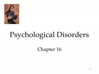
Vignette A 23 yo med student with a 2 day history of headache, blurred/darker vision/loss of vivid color. There is accompanying left retro- orbital pain which worsens with eye movement. She has never had anything like this before, and past medical history is negative. • Anatomical localization?
Questions • Exam: L eye 20/100; R 20/20; L disc is edematous; pupils are equal but when light is shined into L eye pupils are 4mm; and 2 mm when shined into R eye. R gaze: L eye adducts more slowly than the R eye. L gaze eyes are conjugate. Enlarged blind spot noted on the L. • Does the examination refute or support localization? • Cause for visual impairment
Questions • Visual acuity • Light reaction • Eye movements • Visual fields • Cause of visual impairment • Cause of eye movement difficulties
Questions • ROS • PMHx • Tests • Diagnosis • Certainty and how is diagnosis is made • What is the outlook? • What do you disclose?
MRI Scans of the Brain of a 25-Year-Old Woman with Relapsing–Remitting Multiple Sclerosis. An axial FLAIR (fluid-attenuated inversion recovery) image shows multiple ovoid and confluent hyperintense lesions in the periventricular white matter (Panel A). Nine months later, the number and size of the lesions have substantially increased (Panel B). After the administration of gadolinium, many of the lesions demonstrate ring or peripheral enhancement, indicating the breakdown of the blood–brain barrier (Panel C). In Panel D, a parasagittal T1-weighted MRI scan shows multiple regions in which the signal is diminished (referred to as "black holes") in the periventricular white matter and corpus callosum. These regions correspond to the chronic lesions of multiple sclerosis.
Multiple sclerosis: This 35-year-old woman with a history of migraine headaches presented with a two-week history of slurred speech and trouble walking. Her examination was significant for slight left hemiparesis, brisk jaw jerk and bilateral hyperreflexia. Laboratory data demonstrated oligoclonal banding in the cerebrospinal fluid. These FLAIR-weighted axial MR images show multiple high signal lesions within the periventricular white matter. On the sagittal image on the right the signals emanate radially from the corpus callosum.
Relative afferent pupillary defect This is a common case in pupillary examination. Always suspect this if there is no anisocoria. The direct and consensual pupillary responses to light are normal. The swinging light test shows abnormal light response of the affected eye (initial dilatation followed by constriction). For example, if the left eye were abnormal, both pupils constrict when the light is shown into the right eye. When the light is swung to the left eye, both pupils dilate. When the light is swung back to the right eye both pupils again constrict. This reaction indicates a defect in the afferent pupillary fibres from the left eye. The near reflex is normal. Further examination: tell the examiner that you would like to examine the fundus of the affected eye. The most common physical signs would be optic atrophy. Other possibilities include advanced glaucoma, retinitis pigmentosa, old central retinal artery or vein occlusion. The arrows represent the light. A patient with a left relative afferent pupillary defect.
Fig.1 A patient with a right dilated and unreactive pupil. The swinging flash test shows abnormality of the left eye. (Note the dilatation of the left eye when the light is swung to the left.) Fig. 2 A patient with a right dilated and unreactive pupil. The swinging flash test shows abnormality of the right eye (Note dilatation of the left eye when the light is swung to the right).
Left internuclear ophthalmoplegia L R The most common scenario in the examination is young female with history of multiple sclerosis. However, it can also be seen in older patients with cerebrovascular accident. The main feature of this condition is impaired adduction. A favourite question is the site and side of the lesion (see question below). In unilateral case, the affected eye shows failure (or impaired) adduction (failure of conjugate eye movement). The abducting eye shows jerk nystagmus with the quick phase towards the opposite side (this is called ataxic nystagmus but may not be obvious and can be absent). The horizontal saccade is abnormal with the affected eye lagging behind the normal eye. The vertical saccade and convergence are normal.
Internuclear ophthalmoplegia (INO) a. Normal primary position b. Left impaired adductionn on right gaze d. Normal convergence c. Normal left abduction on left gaze
Impact on Visual Fields • Left optic nerve lesion • Optic chiasmal lesion • Left temporal lobe lesion • Left occipital lobe lesion
Central scotoma resulting from inflammation of the optic disc • 2. Junctional scotoma • 3. Bitemporal hemianopia resulting from a lesion around the optic chiasm • 4. Incongruous homonymous hemianopia resulting from a lesion in the optic tract • 5. Homonymous quadrinopia resulting from a lesion in the temporal lobe • 6. Homonymous hemianopia resulting from a lesion in the occipital lobe.
Central scotoma resulting from • inflammation of the optic disc
3. Bitemporal hemianopia resulting from lesion around the optic chiasm There is bitemporal hemianopia which obeys the midline.and suggests a lesion in the optic chiasm. The hemianopia may be subtle and revealed only by comparing two red objects in each hemifield. The red color in the temporal field appears washed out. If the hemianopia does not obey midline consider pseudo-bitemporal hemianopia such as bilateral sectorial retinitis pigmentosa, tilted discs or bilateral inferotemopral retinoschisis. Examine the fundi for any such changes. Features of pituitary abnormalities such as acromegaly, pan-hypopituitarism (smooth skin and absence body hair in male) Look for any scar suggestive of pituitary operation.
4. Incongruous homonymous hemianopia resulting from lesion in the optic tract
5. Homonymous quadrinopia from lesion in the temporal lobe Left superior homonymous quadrinopia Left inferior homonymous quadrinopia
If the patient has a right inferior quadrinopia, you should consider Gertmann's syndrome if the lesion is in their dominant parietal lobe. What is Gertmann's syndrome? Failure of the person to calculate (acalculia), name fingers (finger agnosia), and to tell right from left In patients with hemianopia or inferior quadrinopia, mention you like to perform the optico-kinetic drum test for evidence of parietal lobe lesion. What abnormality may you see? Impaired pursuit to the side of the parietal lobe lesion. Abnormal opticokinetic nystagmus with small saccadic movement replacement the smooth pursuit movement. Normal opticokinetic nystagmus with smooth pursuit movement.
Which other higher sensory perceptions are impaired Astereoagnosia (failure to tell an object through touch) Joint position sense loss and loss of two point discrimination How do you test for sensory inattention? With the patient’s eyes closed alternatively touch the patient’s right then left hand (to ascertain that primary sensory function is intact) and then touch both hands simultaneously. If there is sensory inattention (neglect) the individual will only notice the touch on one side when touched simultaneously.
Impact on a Rightward Saccade • Left middle cerebral artery infarct • Left frontal lobe seizure • Right pontine infarct • Left medial longitudinal fasisculus lacune • Left diabetic third nerve palsy
Movement Definition Associated structure Saccade Pursuit Oculovestibular Convergence
Saccade • Pursuit Function: to keep the fovea on a small moving target, e.g. tracking a tennis ball.As in saccades, pointing the fovea at this moving ball is a 2D problem. • Vestibular ocular reflex/ Oculovestibular reflexFunction: to keep the image of the world stationary on the retina when the head rotates. Try shaking your head while reading this sentence. The fact that you can do this means that your VOR is working, it is keeping your eye still in space.It does this by rotating the eyes in the opposite direction of the head. • Vergence/convergenceFunction: to align a near target on the fovea of each eye.
Vertical Saccade • Vertical saccades originate from burst neurons in the rostral interstitial nucleus of the medial longitudinal fasciculus (riMLF). • The riMLF, like PPRF, produces the phasic command. • Neurons in the interstitial nucleus (INC) convert this to a tonic command (like the pph for horizontal saccades). • The phasic and tonic commands combine in the MN's in III & IV n. • Both sides of the brainstem have neurons that generate upward saccades and others that generate downward saccades. • Do neurons on each side do the same thing; i.e. is this a redundant representation?
Horizontal saccades • Saccades are one of 5 types of eye movements. They are used to point your fovea quickly from one object of interest to another, such as the words of this sentence. • The command for a saccade begins in a structure called the Paramadian Pontine Reticular Formation; the PPRF • Burst neurons in the PPRF generate phasic movement command which is proportional to velocity • Tonic neurons in prepositus hypoglossi (PPH) • converts the phasic command to a tonic command • this is like an integrator which converts velocity to position • Motorneurons (MN's) combine phasic and tonic commands • this contracts muscles • quickly rotates the eyes (phasic component) • & then holds (tonic component) them there against the elastic restoring forces.
III IV V1 V2 VI Contents of the cavernous sinus (red) A. Carotid ArteryB. Trochlear Nerve IVC. Maxillary Nerve V2D. Abducens Nerve VIE. Sphenoid SinusF. Pituitary GlandG. Cavernous SinusH. Ophthalmic Nerve V1I. Oculomotor Nerve III
- Horner's syndrome + cranial nerve 3 and/or 4 and/or 6 and/or V1/V2 dysfunction (and not affecting V3) => suspect cavernous sinus pathology - Horner's syndrome + cranial nerves 3 and/or 4 and/or 6 and/or V1 (and not affecting V2 and V3) dysfunction => suspect superior orbital fissure pathology - Horner's syndrome + cranial nerves 2 (optic nerve), 3, and/or 4 and/or 6 and V1 (and not affecting V2 and V3) dysfunction => suspect orbital apex pathology - Horner's syndrome + optic nerve II dysfunction +/- incomplete cranial nerve 3 dysfunction (and not affecting cranial nerves 3, 4 and 6 and V2 and V3) => suspect posterior orbit pathology - all patients with an asymptomatic, unexplained Horner's syndrome (especially if they have ipsilateral anhidrosis of the face and neck, which implies a preganglionic Horner's syndrome), who are going to be discharged from the ED for pre-arranged follow-up as an outpatient, should have a chest X-ray performed prior to ED discharge - to exclude a mediastinal or apical lung tumor (Pancoast's tumor) or thoracic aneurysm affecting the second neuron
Structural Abnormality Orbit Superior orbital fissure Cavernous sinus Upper brainstem
Autonomic Pupillary Innervation • Sympathetic (Horner syndrome) • Parasympathetic (IIIrd nerve)
For patients with a normal light reaction and whose anisocoria is greatest in the dark => the smaller pupil in the dark is the abnormal pupil (dilation problem with the smaller pupil) Differentiation between physiological anisocoria and Horner's syndrome
Unexplained unilateral Horner's syndrome + face/head pain is a carotid artery dissection until proved otherwise Right Horner’s syndrome: Ptosis Miosis Anhydrosis Vasodilatation
Clinical Pearls - if there is a ptosis of the eyelid on the side of the small pupil => the patient has a Horner's syndrome on that side - if there is a ptosis of the eyelid on the side of the large pupil => the patient has a partial third nerve lesion on that side - with a lesion in the region of the cavernous sinus, there may actually be a reversal of the anisocoria in going from dark to light as a result of unilateral involvement of both parasympathetic and sympathetic axons - a patient with a very small unilateral miotic pupil, that does not decrease in size in response to direct bright light, or dilate in response to dim light => probably has unilateral pharmacolgical miosis secondary to a cholinergic glaucoma drug (eg. pilocarpine) or an anti-cholinesterase agent ("flea collar" anisocoria) - a mydriatic pupil that does not respond to light may appear to be due to 3rd cranial nerve pathology, but it could just be a tonic pupil - the only difference between the two may be that the tonic pupil does eventually constrict slowly on prolonged near fixation - a patient with uncal herniation causing a compressive 3rd cranial nerve palsy, always has some degree of impairment of LOC and is never fully alert - a patient with "apparent" physiological anisocoria may have simple anisocoria secondary to the effect of certain drugs eg. pseudo-ephedrine or serotonin reuptake inhibitors, and the anisocoria (like physiological anisocoria) is also eliminated by the instillation of cocaine eyedrops - blindness in one eye (even if total) never causes anisocoria - retinal pathology (even if very severe) never causes anisocoria
For patients with an abnormal light reaction and whose anisocoria is greatest in bright light conditions => the larger pupil is the abnormal pupil (constriction problem with the larger pupil)