Download
1 / 119
1.21k likes | 1.42k Vues
Topic 1 Cell-cell contacts. Debbie McKenzie. Contact Information Email: debbie.mckenzie@ualberta.ca Office: 1-30 Environmental Engineering Building Access is restricted Please make an appointment. Dates to Remember. Topic Outline. Polarity of cells Extracellular Matrix Function

E N D
Topic 1Cell-cell contacts Debbie McKenzie
Contact Information • Email: debbie.mckenzie@ualberta.ca • Office: 1-30 Environmental Engineering Building • Access is restricted • Please make an appointment
Topic Outline • Polarity of cells • Extracellular Matrix • Function • Structure • Cell Junctions • Functions • Structures • Plant Cell Walls
Cell to Cell Contacts • Concepts in cell biology • Individual cells • DNA replication • RNA transcription • Protein translation and processing • Organisms—multi-cellular • Cellstissuesorgansorganisms • How are cells connected? • How do they communicate?
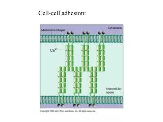
2 extremes of cell-cell adhesion • Connective tissue • Extracellular matrix (ECM) is plentiful • Cells sparse • Matrix bears most of the stress • Cells attached to ECM but few cell-cell contacts
Epithelial cells • Line all cavities and surfaces of the body • Cells tightly bound together in sheets • ECM scanty • Cells bear most of the mechanical stress
What is the ECM? • Tissues not completely composed of cells • Substantial portion is extracellular space • Filled by network of macromolecules • Major component of skin and bones • Minor component of brain, spinal cord
Different types of ECM • Calcified to make bone and teeth • Transparent = cornea • Ropelike = tendons
Components of the ECM • Macromolecules in ECM produced locally • Connective tissue • Fibroblasts • In specialized tissue, components secreted by specialized fibroblasts • Chondroblasts—cartilage • Osteoblasts—bone https://encrypted-tbn3.gstatic.com/images?q=tbn:ANd9GcRGKMKGW-E2Zf3FQl8Tyt--bunKx6Hv-WkOQGsVxDTPx2o-ePjofw
Cartilage http://www.bidmc.org/Research/Departments/Radiology/Laboratories/~/media/Images/CentersandDepartments/Radiology/Research/cartilage/cartilage_with_labels.ashx
Major Components • Glycoaminoglycans (GAGS) • Fibrous proteins • Two types • Mainly structural • Collagen and elastin • Mainly adhesive • Fibronectin and laminin
GAGS • Long polysaccharide chains • Usually covalently cross-linked to protein • Proteoglycans • GAGs/proteoglycans form highly hydrated gel-like substance • Fibrous proteins embedded in the gel • Permits rapid diffusion of nutrients, metabolites and hormones
GAGS • Comprised of repeating disaccharide units • 1 of 2 sugar groups is always an amino sugar • N-acetylglucosamine • N-acetylgalactosamine • 2nd sugar • Usually uronic acid • Glucuronic or iduronic • Highly negatively charged
4 major groups of GAGS • Hyaluronan • Simplest GAG • Facilitates cell migration during tissue morphogenesis and repair
Chondroitin sulfate/dermatan sulfate • Heparan sulfate and heparin • Keratin sulfate • All are sulfated GAGS • Covalently attached to protein • Core protein is usually glycoprotein
Proteoglycans • Can regulate activity of signalling cells • GAG chains form gels of varying pore size and density • Sieving activity • i.e., heparan sulfate proteoglycan—perlecan • Sieving function in basal lamina of kidney glomerulus
Protein binding to a proteoglycan • Controls activity of the protein by: • Immobilizing the protein close to where it was produced • Sterically block its activity • Provide a reservoir for delayed release • Alter or concentrate the protein for more effective presentation to cell surface receptors
Collagen • Major protein of ECM • 25% of total protein mass in mammals • Long, stiff triple-stranded helical structure • 3 alpha helices wound around each other • Rich in proline and glycine
Collagen https://encrypted-tbn2.gstatic.com/images?q=tbn:ANd9GcQp02fBM7aNTHthagYB2ropWBixh719aH-X1jVABhQI0DL3EYvA-[[
Collagen Synthesis • Specific proline and lysine aas are hydroxylated in the ER • Secreted with non-helical extensions on each end • “propeptides” • Propeptides removed in extracellular space
More on Collagen • ~25 different collagens • Encoded by different genes • 5 main types in connective tissue
Scurvy • Vitamin C deficiency • Impacts hydroxylation of proline • Without hydroxylated proline • Can’t form stable triple helix of collagen • Collagen degraded • With time, loss of pre-existing collagen in ECM • Blood vessels become fragile, teeth loose
Other collagen-related diseases • Many steps to collagen fibril synthesis • Many places for “failure” • Mutations in type I collagen • Osteogenesisimperfecta • Weak bones • Mutations in type II collagen • Chondrodysplasias • Abnormal cartilage • Bone and joint abnormalities
Fibril-associated collagens • Help organize fibrils • Types IX and XII • Triple helix interrupted by one or two short non-helical regions • More flexible • Retain propeptides • Mediate the organization of the collagen fibrils
Collagen Fibril Organization • Mammalian skin • Wickerwork pattern of collagen fibrils • Tendons • Collagen fibrils arranged in parallel bundles • Mature bone and cornea • Plywood like layers of collagen fibrils
Elastin • Many vertebrate tissues need to be strong and elastic • Contain network of elastic fibers • Main component is elastin • Rich in proline and glycine • Little/no hydroxyproline or hydroxylysine • Highly cross-linked to each other • Elastin core covered with microfibrils of fibrillin
Adhesive Glycoproteins anchor cells to ECM • Two most common forms • Fibronectin • Laminins • Members of the integrin family
Fibronectin • Widely distributed in vertebrates • Can be • Soluble (blood) • Insoluble (ECM) • Intermediate form (associated with cell surfaces
Fibronectin Structure • Dimer of 2 large subunits connected by pair of disulfide bonds • 1 domain binds heparin • 1 domain binds collagen
Fibronectin Function • Bridging molecule between ECM and cells • In vitro • Cells in culture dish coated with fibronectin attach more readily to surface • Involved in cellular movement • Serve as guides for migrating cells
Laminins • Found primarily in basal lamina • Underlies epithelial cells separating them from the connective tissue • Basal lamina • Serves as structural support • Permeability barrier • Contains type IV collagen, proteoglycans, lamins and entactin
Organization of Basal Lamina • Laminins localized on surface of lamina that faces epithelial cells • Fibronectin anchor cells of the connective tissue
Altering the basal lamina • Matrix metalloproteases • Use metal ions as cofactors • Can locally degrade the ECM • Allows cells to pass through • i.e., leukocytes migrating to injured tissues
Properties of Laminin • Huge protein (850 kDa) • 3 polypeptides (several types of each)
Integrins • Large family of receptors • Binds fibronectin, laminin etc • Two subunit • Different binding specificities
Integrin Function • Regulate cell movement and attachment • Also interact with intracellular signalling pathways • Binding to growth factors that activate MAP kinases • Inside out signalling—internal changes in cell affect integrins on the surface
Integrins and Cytoskeleton • Link ECM and cytoskeleton indirectly • Migratory and non-epithelial cells attach via focal adhesions • Epithelial cells attach via hemidesmosomes
Cell Junctions • Responsible for cell to cell connections • 3 functional classifications • Occluding junctions (tight junctions) • Anchoring junctions • Via actin filament attachment sites or intermediate filament attachment sites • Communicating junctions • Electrical or chemical signalling
Tight Junctions • Example: epithelium of mammalian small intestine • Epithelial cells regulate transport of materials from lumen of the gut into the extracellular fluid on the other side of the cell • Don’t want materials leaking in between the cells • Tight junctions
Experimental Evidence • Use of tracer dyes • Tracers can’t traverse between the cells