Download

1 / 74
780 likes | 1.07k Vues
Clinical features and research opportunities in rheumatoid arthritis. Clinical Immunology March 26, 2013. HARVARD MEDICAL SCHOOL. Overview. Clinical characteristics and pathophysiology Differential diagnosis Exam and laboratory studies Treatment strategy Research opportunities. Overview.

E N D
Clinical features and research opportunities in rheumatoid arthritis Clinical Immunology March 26, 2013 HARVARD MEDICAL SCHOOL
Overview • Clinical characteristics and pathophysiology • Differential diagnosis • Exam and laboratory studies • Treatment strategy • Research opportunities
Overview • Clinical characteristics and pathophysiology • Differential diagnosis • Exam and laboratory studies • Treatment strategy • Research opportunities
Rheumatoid Synovium Normal Synovium Lining Sublining
The 1-2-3 of Rheumatoid Arthritis Lee, Kiener and Brenner, Synoviocytes 2004
Understanding pathogenesis Klareskog et al Lancet 2009
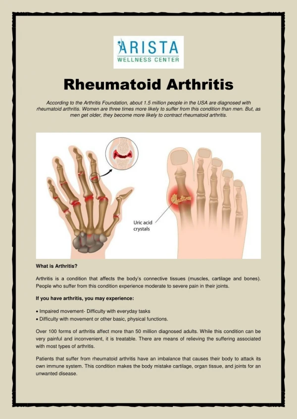
Clinical characteristics • Systemic chronic inflammatory disease • Mainly affects synovial joints • Variable expression • Extra-articular manifestations (e.g., nodules, ILD, ocular) • Prevalence ~1% • Worldwide distribution • Female: Male ratio 3:1 • Peak age of onset 30 – 50 years (median in 40’s)
ACR Criteria for Diagnosis • Four or more of the following criteria must be present: • Morning stiffness >1 hour • Arthritis of >3 joint areas • Arthritis of hand joints (MCPs, PIPs, wrists) • Symmetric swelling (arthritis) • Serum rheumatoid factor • Rheumatoid nodules • Radiographic changes First four criteria must be present for 6 weeks or more
Overview • Clinical characteristics and pathophysiology • Differential diagnosis • Exam and laboratory studies • Treatment strategy • Research opportunities
Rheumatoid Arthritis Psoriatic Arthritis Inflammatory bowel disease Ankylosing spondylitis Crystal – Gout, Pseudogout SLE, Vasculitis PMR-GCA Any “immune complex” illness Paraneoplastic syndrome Viral – Parvovirus, HepBSAg, HCV, Rubella Bacterial – Lyme, GC, chlamydia Osteoarthritis, bursitis, tendonitis Differential Diagnosis
Conceptual organization • Inflammatory vs. non-inflammatory • synovitis vs structural • Articular vs. non-articular • Systemic vs. regional • Polyarticular vs. monarticular • Extra-articular manifestations Note: Older patients need more careful history and physical exam-labs often confusing
Pertinent historical features • Duration • acute vs chronic • gradual vs abrupt onset • Pattern • symmetrical vs asymetrical • large vs small joints • morning stiffness • effect of activity • Joint distribution • DIP vs PIP/MCP DIP PIP MCP
Overview • Clinical characteristics and pathophysiology • Differential diagnosis • Exam and laboratory studies • Treatment strategy • Research opportunities
Physical exam of joint • Tenderness • synovitis = tender joint • mechanical or periarticular lesions (bursitis and tendonitis) = tenderness often localized • Swelling • bony vs. soft tissue swelling • Pattern • proximal vs. distal • asymetric vs. symmetric • DIP and nail changes
Proximal InterPhalangeal joint • Swelling is confined to the area of the joint capsule • Synovial thickening feels like a firm sponge PIP
Rheumatoid Arthritis Psoriatic Arthritis Inflammatory bowel disease Ankylosing spondylitis Crystal – Gout, Pseudogout SLE, Vasculitis PMR-GCA Any “immune complex” illness Paraneoplastic syndrome Viral – Parvovirus, HepBSAg, HCV, Rubella Bacterial – Lyme, GC, chlamydia Osteoarthritis, bursitis, tendonitis Laboratory values based on DiffDx
Rheumatoid Arthritis Psoriatic Arthritis Inflammatory bowel disease Ankylosing spondylitis Crystal – Gout, Pseudogout SLE, Vasculitis PMR-GCA Laboratory values based on DiffDx • Markers of inflammation • ESR and CRP • Auto-antibodies • RF and CCP • X-rays hands/feet • erosions
Rheumatoid Arthritis Psoriatic Arthritis Inflammatory bowel disease Ankylosing spondylitis Crystal – Gout, Pseudogout SLE, Vasculitis PMR-GCA Laboratory values based on DiffDx • joint aspiration • Presence of crystals • blood • Uric acid • X-rays • erosions
Rheumatoid Arthritis Psoriatic Arthritis Inflammatory bowel disease Ankylosing spondylitis Crystal – Gout, Pseudogout SLE, Vasculitis PMR-GCA Laboratory values based on DiffDx • autoantibodies • ANA • ANCA • blood • CH50 • urine • urinalysis • X-rays • lack of erosions
Joint fluid analysis • Cell count and differential • Crystals • Gram stain and culture non-inflammatory <1500 mildly inflammatory 1500-3500 inflammatory >3500 possible infection >50,000
Clinical utility of x-rays • X-rays show only bone, not cartilage or synovium • Lesions must correlate w/ clinical picture • Erosive pattern (or lack) useful in diff. diagnosis • Early inflammatory lesions often non-specific • X-ray changes take months to occur • avascular necrosis not visible for 6 wks • spondylitis not evident for 2 – 10 yrs • Valuable for plotting the clinical course in terms of structural changes
Patterns of radiographic changes RA OA gout
Patterns of radiographic changes psoriasis RA OA gout CPPD http://www.gentili.net/Hand/summary.htm
Progression of RA erosions How fast is joint damage progressing? • Soft-tissue swelling, osteopenia, no erosions • Thinning of cortex with minimal joint space narrowing C. Marginal erosion with joint space narrowing A B C ACR Clinical Slide Collection, 1997.
Overview • Clinical characteristics and pathophysiology • Differential diagnosis • Exam and laboratory studies • Treatment strategy • Research opportunities
Typical Course • Damage occurs early in most patients • 2 yrs: 50% show joint space narrowing or erosions • 10 yrs: 50% of young working patients are disabled • Death comes early • Multiple causes (especially cardiovascular) • Women lose 10 yrs, men lose 4 yrs Pincus, et al. Rheum Dis Clin North Am. 1993;19:123–151.
Treatment principles • Determine spectrum of disease • Use the safest treatment plan that matches the aggressiveness of the disease • Monitor treatment for adverse effects • Monitor disease activity, revise Rx as needed
abatacept (Orencia)
Overview • Clinical characteristics and pathophysiology • Differential diagnosis • Exam and laboratory studies • Treatment strategy • Research opportunities
Cost is increasing, productivity is decreasing We need new drugs to treat RA and other complex traits! Scannell et al Nat Rev Drug Discovery (2012)
Major driver of cost is failure in clinical trials… …and most drugs fail due to lack of efficacy or toxicity in humans target Medicinal chemistry trials
“Target validation” is key to avoid failure from efficacy/safety Current models are ineffective at choosing targets that are safe and effective in humans target Medicinal chemistry trials
target Medicinal chemistry trials We determine dose-response in clinical trials, after many years and millions of dollars
target Medicinal chemistry trials We aspire to determine dose-response at the time of target validation
Human genetics is a unique tool for target validation • Nature’s perturbation of many drug targets in the human genome • Links physiological state in humans (e.g., disease risk) to a target perturbation • Indicates gain- or loss-of-function • Provides allelic series for range of effect on perturbing a potential drug target Dose-response curves derived from human genetics
The history and success of GWAS – illuminating for common phenotypes 2007 Data: www.genome.gov/GWAStudies - slide from Sara Pulit and Paul de Bakker
The history and success of GWAS – illuminating for common phenotypes 2008 Data: www.genome.gov/GWAStudies - slide from Sara Pulit and Paul de Bakker
The history and success of GWAS – illuminating for common phenotypes 2009 Data: www.genome.gov/GWAStudies - slide from Sara Pulit and Paul de Bakker
The history and success of GWAS – illuminating for common phenotypes 2010 Data: www.genome.gov/GWAStudies - slide from Sara Pulit and Paul de Bakker
The history and success of GWAS – illuminating for common phenotypes 2011 Data: www.genome.gov/GWAStudies - slide from Sara Pulit and Paul de Bakker
The history and success of GWAS – illuminating for common phenotypes 2012 Data: www.genome.gov/GWAStudies - slide from Sara Pulit and Paul de Bakker
Similarly, great success in unraveling genetics of RA GWAS 1,522 RA cases, 1,850 controls No. GWAS hits = 3 Total No. risk loci = 5* (* includes replication beyond GWAS) 15 10 5 0 Chromosomal position Plenge et al NEJM 2007