Download
1 / 149
1.59k likes | 2.89k Vues
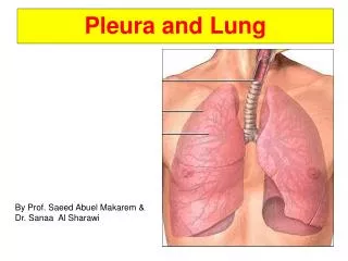
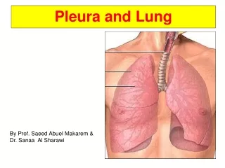
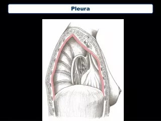
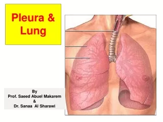
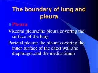
The boundary of lung and pleura. Pleura Visceral pleura:the pleura covering the surface of the lung Parietal pleura: the pleura covering the inner surface of the chest wall,the diaphragm,and the mediastinum.

E N D
The boundary of lung and pleura • Pleura Visceral pleura:the pleura covering the surface of the lung Parietal pleura: the pleura covering the inner surface of the chest wall,the diaphragm,and the mediastinum
On the right, the dome of the diaphragm is situated at a level approximating the fifth rib or fifth interspace at the midclavicular line.The dome of the left diaphragm is ordinarily about 1 inch lower than the right.
INSPECTION Inspection of the chest,productive of the maximum amount of information, requires the following: • 1. First and foremost,a definite desire to see and to appreciate every visible abnormality • 2. The patient stripped to the waist • 3. Good lighting
INSPECTION • 4. A thorough knowledge of topographic anatomy • 5. The examiner and patient in a comfortable position throughout the examination. If either the physician or patient is uncomfortable,the examination may be hurried and consequently less thorough. It is important that the patient be absolutely straight,whether seated or supine.
INSPECTION Normal thorax You should know that in normal subjects there is a wide variation in the size and shape of the thorax.At times it is difficult to be certain where the normal variations and definite pathologic changes begin.
INSPECTION Normal thorax The anteroposterior diameter of the thorax in the normal adult is less than the transverse diameter.
INSPECTION what to observe • 1.First: the general nutrition and musculoskeletal development 2.Next: the skin and breasts • 3.vein and subcutaneous emphysema
INSPECTION • 4.the anteroposterior diameter of the thorax persons with pulmonary emphysema --barrel chest • 5.the general slope of the ribs normal : 45 º degree angle patients with emphysema :the ribs are nearly horizontal ; this angle becomes abnormally wide
INSPECTION • 6.retraction or bulging of interspaces • Retraction of the interspaces:obstruction of the respiratory tract • Bulging of interspaces :a massive pleural effusion,tension pneumothorax
INSPECTION • 7.the rate and depth of quiet breathing • in the adult at rest the normal respiratory rate is approximately 16 to 18 breaths per minute and is quite regular in depth and rhythm • increase in the respiratory rate :fever
INSPECTION • 8.Alterations in shape of the thorax • In the normal subject,the two sides of the chest move synchronously and expand equally • Unilateral retraction of the thorax :a thickened fibrotic pleura • Pigeon chest • Funnel chest
INSPECTION • 9.Types of respiration • (1)Dyspnea :difficulty in breathing ; participation of the accessory respiratory muscles • Inspiratory dyspnea :obstruction of the trachea or major bronchi (tumor,laryngitis) • Expiratory dyspnea :obstruction in the bronchioles and smaller bronchi (asthma)
INSPECTION • 9.Types of respiration • (2)Bradypnea : abnormal slowing of respiration • (3)Apnea : temporary cessation of breathing • (4)Tachypnea : increased respiratory rate • (5)Hyperpnea : an increase in thedepth of respiration • (6)Hyperventilation :an abnormal increase in both rate and depth of respiration(it is seen in diabetic acidosis and highly emotional states)
INSPECTION • 9.Types of respiration • (7) restrained breathing :the inspiratory phase is suddenly interrupted as a result of pain associated with acute pleuritis ; The respirations are quite shallow but more rapid than normal
INSPECTION • 9.Types of respiration • (8)tidal respiration :is characterized by periods of rapidly increasing rate and depth of respiration, which within a matter of a few more respiratory cycles becomes shallower and shallower until respiration ceases.This is followed by a period of apnea,which may last a few seconds to as long as 30 seconds.periodic respiration may be present in many relatively severe disease states.
INSPECTION • 9.Types of respiration • (9)Sighing respiration :occurs when the normal respiratory rhythm is interrupted by a deep inspiration,which is followed by a prolonged expiration and ordinarily is accompanied by audible sighing. it is rarely associated with organic disease;instead it is almost always a manifestation of emotional tension.
INSPECTION • 9.Types of respiration • (10)Ataxic breathing: is characterized by unpredictable irregularity . Breaths may be shallow or deep,and stop for short periods.
PALPATION Thoracic expansion • Variations in expansion are more readily detectable on the anterior surface where there is greater range of motion. • The examiner's hands should be placed over the lower anterolateral aspect of the chest. • Expansion should be tested during both quiet and deep inspiration.
PALPATION Thoracic expansion • Expansion may be limited as the result of acute pleurisy,fibrous thickening of the pleura (fibrothorax),fractured ribs,or other trauma to the chest wall.
PALPATION Fremitus • Vocal fremitus :Vocal fremitus is a palpable vibration of the thoracic wall produced by phonation .
PALPATION Vocal fremitus: The sounds that arise in the larynx are transmitted down along the air column of the tracheobronchoalveolar system into the bronchi of each lung,on through the smaller bronchi into the alveoli,setting in motion the thoracic wall that acts as a large resonator. Thus,vibrations are produced in the chest wall that can be felt by the hand of the examiner.
PALPATION Vocal fremitus: In eliciting vocal fremitus the patient is directed to count “one,two,three”---“one,two,three”,to repeat the words“ninety-nine”—“ninety-nine”,or to say “ e-e-e,e-e-e,e-e-e”. The patient should speak with a voice of uniform intensity throughout the examination so that the examiner can better compare the transmission of the fremitus in different areas of the chest.
PALPATION Vocal fremitus: • The vocal fremitus is perceived by placing the palmar aspect of the fingers or ulnar aspect of the hand against the chest wall.Usually both hands are used,placing them in corresponding areas so that comparison of the two sides can be made. If only one hand is used,it should be moved from one place to the corresponding area of the other side to compare the transmission of sound.
PALPATION • Normal variations of vocal fremitus. • The intensity of the vocal fremitus perceived in the normal subject is governed by the following: 1.Intensity of the voice 2.Pitch of the voice 3.Varying relations of the bronchi to the chest wall 4.Varying thickness of the thoracic wall
PALPATION • In general,vocal fremitus is most prominent in the regions of the thorax where the large bronchi are the closest to the thoracic wall and tends to become less intense as one progresses farther from the major bronchi.In the normal person the fremitus is found at maximum intensity over the upper thorax both anteriorly and posteriorly.It is least intense at the bases.
PALPATION • Also the intensity of the fremitus will vary with the thickness of the thoracic wall.In a thin person the vibrations will be more intense than in the normally developed or obese patient. There is considerable variation from patient to patient.
PALPATION • Alternations of vocal fremitus • increased vocal fremitus ----consolidation of the lungs :lobar pneumonia • Decreased fremitus ----fibrous thickening of the pleura:fluid in the pleural space or pneumothorax • absent fremitus ----major bronchus is obstructed :tumor
PALPATION • pleural friction fremitus:As the result of acute pleurisy,the inflamed pleural surfaces rub against one another,producing a pleural friction rub that may be detected by the examining hand.
PALPATION • pleural friction fremitus • When present,it is palpable usually in both phases of respiration. • Friction rubs most commonly are felt as well as heard in the inferior anterolateral portion of the chest,the area of greatest thoracic excursion.
PALPATION • Crepitation • Crepitation may be palpated when the sub cutaneous tissues contain fine beads of air. • This condition is known as subcutaneous emphysema. • A somewhat similar sensation can be produced by rolling a lock of hair between the thumb and fingers.
PERCUSSION There are two principal methods that may be used for percussion of the thorax, abdomen,or other structures.
PERCUSSION 1. Mediate percussion is that in which the examiner strikes the middle finger of one hand held against the thorax, thus producing a sound by setting the chest wall and underlying structures in motion. This is the method in almost universal use today.
PERCUSSION 2. Immediate percussion may be useful in demonstrating changes in percussion note.This can be done by striking the chest with the tips of all of the fingers held firmly together.
PERCUSSION Practical experience has demonstrated that useful sounds produced by percussion probably do not penetrate more than about 4 to 5cm below the surface. Also a lesion must be at least 2 or 3cm in diameter to be detectable. Thus,it is obvious that percussion will only locate rather gross abnormalities.
PERCUSSION • To obtain the maximum information from percussion: 1. The distal phalanx of the pleximeter finger must be pressed firmly on the chest wall;otherwise,a clear note is not ob tained. 2. The plexor finger should strike the pleximeter finger only instantaneously and must be immediately withdrawn.
PERCUSSION • Usually percussion is performed above the clavicles in the supraclavicular spaces and downward.Next,each lateral wall is examined, beginning in the axilla and working down to the coastal margin. With the pleximeter finger always parallel to the ribs--never cross them.
PERCUSSION • In examining the back of the chest the patient should have his head inclined forward and the forearms crossed comfortably at the waist to move the scapulae as far laterally as possible.
PERCUSSION • Examination is started at the apices, where the percussion note as well as the width of the isthmus of normal resonance over the apex is determined . Bounded medially by the neck muscles and laterally by the shoulder girdle,this band of resonance is normally about 5 cm wide.
PERCUSSION • The percussion is continued downward, interspace by interspace,to the bases where the location and range of motion of each hemidiaphragm is ascertained.
PERCUSSION • Analysis of percussion tones The sound waves produced by percussion are influenced more by the character of the immediate underlying structures than by those more distant. Consequently the tone produced by percussion over the airfilled lung will be different from the tone heard over a solid structure,such as the heart or liver.This is the basis for the scientific application of percussion.
PERCUSSION • Percussion sounds • 1. Resonance: the sounds heard normally over lungs • 2. Hyperresonance:The hyperresonant note in the adult is commonly the result of emphysema.
PERCUSSION • Percussion sounds • 3. Tympany : It never occurs in the normal chest,except below the dome of the left hemidiaphragm,where the underlying stomach and bowel will produce tympany.
PERCUSSION • Percussion sounds • 4.Dullness: Dullness tends to occur when there is solid or liquid medium present in the underlying lung in proportion to the amount of air in the lung tissue. Thus,dullness will be found when there is consolidation of lung,such as occurs in pneumonia,or when there is a moderate amount of fluid in the pleural space with some underlying air-containing lung.
PERCUSSION • Percussion sounds • 5. Flatness is the term used to describe the percussion note when resonance is absent. Flatness will be present when there is a very large fluid mass,such as in an extensive pleura1 effusion with little underlying air-bearing lung to influence the sound.
PERCUSSION • Percussion sounds Over the apices,where there are large amounts of muscle and bone with relatively little underlying resonant lung,the note is less resonant than over the bases,where there is a relatively greater amount of lung with less thoracic wall and muscle.
PERCUSSION • Percussion sounds The development of the pectoral muscles,the heavy muscles of the back,the breasts,and the scapulae,all tend to make the percussion note lessresonant (duller).