The Enterobacteriaceae Basic Properties
The Enterobacteriaceae Basic Properties. Dr. John R. Warren Department of Pathology Northwestern University Feinberg School of Medicine June 2007. Characteristics of the Enterobacteriaceae. Gram-negative rods Glucose is fermented with strong acid formation and often gas

The Enterobacteriaceae Basic Properties
E N D
Presentation Transcript
The EnterobacteriaceaeBasic Properties Dr. John R. Warren Department of Pathology Northwestern University Feinberg School of Medicine June 2007
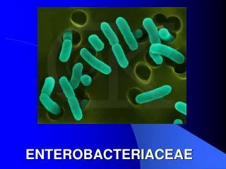
Characteristics of the Enterobacteriaceae • Gram-negative rods • Glucose is fermented with strong acid formation and often gas • Cytochrome oxidase activity is negative • Nitrate is reduced to nitrite
Gram’s Stain for Bacterial Morphology • Crystal violet binds to cell wall peptidoglycan with Gram’s iodine as a mordant • Safranin or basic fuchsin counterstains bacterial cells decolorized by alcohol-acetone
Gram’s Stain for Bacterial Morphology • Thick cell-wall peptidoglycan layer of gram-positive bacteria strongly binds crystal violet and resists decolorization by alcohol-acetone • Thin cell-wall peptidoglycan layer of gram-negative bacteria located beneath a thick lipid-rich outer membrane weakly binds crystal violet that is readily removed by alcohol-acetone decolorization
Gram’s Stain Procedure • Flood surface of smear with crystal violet solution • After 1 min thoroughly rinse with cold tap water • Flood smear with Gram’s iodine for 1 min • Rinse smear with acetone-alcohol decolorizer until no more crystal violet in rinse effluent • Rinse with cold tap water • Flood smear with safranin (regular Gram’s stain) or basic fuchsin (enhanced Gram’s stain) • Rinse with cold tap water • Dry smear in slide rack • Microscopically examine stained smear using oil-immersion light microscopy
Glucose Fermentation • Oxidation-reduction of glucose in the absence of molecular oxygen (anaerobic glycolysis) • Energy from hydrolysis of chemical bonds in anaerobic glycolysis captured as high energy phosphate bonds of adenosine triphosphate (ATP) • NAD is reduced to NADH2 by accepting electrons during glycolytic conversion of glucose to pyruvate • NADH2 in turn reduces pyruvate with oxidation of NADH2 to NAD which supports continued anaerobic glycolysis, and generation from pyruvate of alcohols, carboxylic acids, and CO2 gas • End products of glucose fermentation: organic acids and CO2 gas • Fermentation detected by acidification of glucose-containing broth (color change in broth or agar medium containing pH indicators), and (for aerogenic species) production of gas (fractures in agar, gas bubbles in inverted Durham tube) • pH indicators: phenol red (yellow at acid pH), methyl red (red at acid pH), neutral red (red at acid pH), bromcresol purple (yellow at acid pH)
Spot Cytochrome Oxidase Test • The spot cytochrome oxidase test is the first test performed with gram-negative bacteria recovered in culture • The optimal plate medium for a spot cytochrome oxidase test is a trypticase soy agar (TSA) containing 5% sheep blood • Bacterial colonies should be 18 to 24 hr old
Spot Cytochrome Oxidase Test • In a positive test, bacterial cytochrome oxidase oxidizes the colorless reduced substrate tetramethyl-p-phenylenediamine dihydrochloride (TPDD) forming a dark purple oxidized indophenol product • Streak a small portion of bacterial colony to filter paper soaked with a 1% solution of TPDD • If the streak mark turns purple in 10 sec or less, the spot oxidase test is interpreted as positive
Nitrate Reduction • Enterobacteriaceae extract oxygen from nitrate (NO3)producing nitrite (NO2) • NO2 detected by reaction with α-naphthylamine and sulfanilic acid producing a red colored complex • Absence of red color indicates either no reduction of NO3 or reduction to products other than NO2 (denitrification) • Confirmation of true negative test: addition of zinc ions which reduce NO3 to NO2 producing a red color in the presence of α-naphthylamine and sulfanilic acid
Enterobacteriaceae: Genetic Properties • Chromosomal DNA has 39-59% guanine-plus-cytosine (G+C) content • Escherichia coli is the type genus and species of the Enterobacteriaceae • Species of Enterobacteriaceae more closely related by evolutionary distance to Escherichia coli than to organisms of other families (Pseudomonadaceae, Aeromonadaceae)
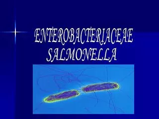
Enterobacteriaceae: Major Genera • Escherichia • Shigella • Salmonella • Edwardsiella • Citrobacter • Yersinia • Klebsiella • Enterobacter • Serratia • Proteus • Morganella • Providencia
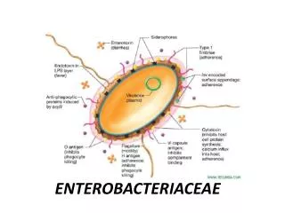
Enterobacteriaceae: Microbiological Properties • Gram-negative and rod shaped (bacilli) • Ferment rather than oxidize D-glucose with acid and (often) gas production • Reduce nitrate to nitrite • Grow readily on 5% sheep blood or chocolate agar in carbon dioxide or ambient air • Grow anaerobically (facultative anaerobes)
Enterobacteriaceae: Microbiological Properties • Catalase positive and cytochrome oxidase negative • Grow readily on MacConkey (MAC) and eosin methylene blue (EMB) agars • Grow readily at 35oC except Yersinia (25o-30oC) • Motile by peritrichous flagella except Shigella and Klebsiella which are non-motile • Do not form spores
Enterobacteriaceae: Natural Habitats • Environmental sites (soil, water, and plants) • Intestines of humans and animals
Enterobacteriaceae: Modes of Infection • Contaminated food and water (Salmonella spp., Shigella spp., Yersinia enterocolitica, Escherichia coli O157:H7) • Endogenous (urinary tract infection, primary bacterial peritonitis, abdominal abscess) • Abnormal host colonization (nosocomial pneumonia) • Transfer between debilitated patients • Insect (flea) vector (unique for Yersiniapestis)
Enterobacteriaceae: Types of Infectious Disease • Intestinal (diarrheal) infection • Extraintestinal infection Urinary tract (primarily cystitis) Respiratory (nosocomial pneumonia) Wound (surgical wound infection) Bloodstream (gram-negative bacteremia) Central nervous system (neonatal meningitis)
Enterobacteriaceae: Urinary Tract Infection, Pneumonia • Urinary tract infection: Escherichia coli, Klebsiella pneumoniae, Enterobacter spp., and Proteus mirabilis • Pneumonia: Enterobacter spp., Klebsiella pneumoniae, Escherichiacoli, and Proteus mirabilis
Enterobacteriaceae: Wound Infection, Bacteremia • Wound Infection: Escherichia coli, Enterobacter spp., Klebsiellapneumoniae, and Proteus mirabilis • Bacteremia: Escherichia coli, Enterobacter spp., Klebsiellapneumoniae, and Proteus mirabilis
Enterobacteriaceae: Nosocomial Infections in the United States 1986-1989 and 1990-19961 • Escherichia coli 27,871 (13.7%) • Enterobacter spp. 12,757 (6.2%) • Klebsiella pneumoniae 11,015 (5.4%) • Proteus mirabilis 4,662 (2.3%) • Serratia marcescens 3,010 (1.5%) • Citrobacter spp. 2,912 (1.4%) 1Enteric Reference Laboratory, Centers for Disease Control and Prevention
Enterobacteriaceae: Intestinal Infection • Shigella sonnei (serogroup D) • Salmonella serotype Enteritidis • Salmonella serotype Typhimurium • Shigella flexneri (serogroup B) • Escherichia coli O157:H7 • Yersinia enterocolitica
Triple Sugar Iron (TSI) Agar • Yeast extract 0.3% (% = grams/100 mL) • Beef extract 0.3% • Peptone 1.5% • Proteose peptone 0.5% Total Protein = 2.6% • Lactose 1.0% • Sucrose1 1.0% • Glucose 0.1% Carbohydrate = 2.1% 1Absent in Kligler Iron Agar
Triple Sugar Iron (TSI) Agar • Ferrous sulfate • Sodium thiosulfate • Sodium chloride • Agar (1.2%) • Phenol red • pH = 7.4
TSI Reactions of the Enterobacteriaceae • Yellow deep, purple slant: acid deep due to glucose fermentation , no lactose or sucrose fermentation with alkaline slant due to production of amine’s from protein • Black deep, purple slant: acid deep due to glucose fermentation with H2S production, no lactose or sucrose fermentation • Yellow deep and slant: acid deep and slant due to glucose as well as lactose and/or sucrose fermentation • Black deep and yellow or black slant: acid deep and slant with glucose and lactose and/or sucrose fermentation with H2S production • Fracturing or lifting of agar from base of culture tube: CO2 production
TSI Reactions of the Enterobacteriaceae • A/A + g = acid/acid plus gas (CO2) • A/A = acid/acid • A/A + g, H2S = acid/acid plus gas, H2S • Alk/A = alkaline/acid • Alk/A + g = alkaline/acid plus gas • Alk/A + g, H2S = alkaline/acid plus gas, H2S • Alk/A + g, H2S (w) = alkaline/acid plus gas, H2S (weak)
A/A + g • Escherichia coli • Klebsiella pneumoniae • Klebsiella oxytoca • Enterobacter aerogenes • Enterobacter cloacae • Serratia marcescens1, 2 1Non-lactose, sucrose fermenter 255% + g
A/A • Serratia marcescens1, 2 • Yersinia enterocolitica2 145% of strains 2Non-lactose, sucrose fermenter
A/A + g, H2S • Citrobacter freundii • Proteus vulgaris1 1Non-lactose, sucrose fermenter
Alk/A • Shigella • Providencia
Alk/A + g • Salmonella serotype Paratyphi A
Alk/A + g, H2S • Salmonella (most serotypes) • Proteus mirabilis • Edwardsiella tarda
Alk/A + g, H2S (w) • Salmonella serotype Typhi
MacConkey (MAC) Agar • Peptone 1.7% • Polypeptone 0.3% • Lactose1 1.0% • Bile salts2 0.15% • Crystal violet2 • Neutral red3 • Sodium chloride 0.5% • Agar 1.35% • pH=7.1 1Differential medium for lactose fermentation 2Inhibit gram positives and fastidious gram-negatives; MAC agar selective for gram-negatives 3Red color at pH < 6.8
Eosin Methylene Blue (EMB) Agar (Levine) • Peptone 1.0% • Lactose1 0.5% • Eosin y2 • Methylene blue2 • Agar • pH = 7.2 1Modified formula also contains sucrose (0.5%) 2Inhibit gram-positives and fastidious gram-negatives; selective for gram-negatives. Eosin y and methylene blue form a precipitate at acid pH; differential for lactose fermentation
Bacterial Utilization of Lactose • Presence of β-galactoside permease: Transport of β-galactoside (lactose) across the bacterial cell wall • Presence of β-galactosidase: Hydrolysis of β-galactoside bond (lactoseglucose + galactose) • ONPG: Orthonitrophenyl-β-D-galacto- pyranoside
Differential Reactions of the Enterobacteriaceae by TSI, ONPG, and MAC • Escherichia coli Red colonies, (A/A, ONPG+) pitted • Klebsiella1 Red colonies, (A/A, ONPG+) mucoid • Enterobacter Red colonies (A/A, ONPG+) • Citrobacter2Red or colorless (A/A or Alk/A, ONPG+) colonies • Serratia Colorless colonies (A/A, ONPG+) 1K. pneumoniae, indole –, K. oxytoca, indole + 2C. freundii, indole – and H2S +, C. koseri, indole + and H2S –
Differential Reactions of the Enterobacteriaceae by TSI, ONPG, and MAC • Shigella Colorless Colonies (Alk/A; ONPG – A, B, and C1; ONPG + D1) • Salmonella Colorless Colonies (Alk/A + H2S; ONPG –) • Proteus Colorless Colonies (Alk/A + H2S2; ONPG –) • Edwardsiella tarda Colorless Colonies (Alk/A + H2S; ONPG–) • Yersinia Colorless Colonies (A/A, ONPG +) 1Shigella A, B, and C, ornithine –; Shigella D, ornithine + 2Proteus mirabilis. P. vulgaris sucrose + with A/A + H2S on TSI
Differential Reactions of the Enterobacteriaceae by EMB • Escherichia coli Colonies with metallic green sheen • Klebsiella Colonies with precipitate (ppt) and mucoid appearance • Enterobacter Colonies with ppt • Citrobacter Colonies with/without ppt • Serratia Colonies without ppt • Shigella Colonies without ppt • Salmonella Colonies without ppt • Proteus Colonies without ppt • Yersinia Colonies without ppt
ONPG Reaction and Lactose Fermentation (Lac) ONPG Lac Escherichia coli + + Shigella sonnei + – Citrobacter + +/– Yersinia enterocolitica + – Klebsiella + + Serratia marcescens + –
Xylose Lysine Deoxycholate (XLD) Agar: Composition • Xylose 0.35% • Lysine 0.5% • Lactose 0.75% • Sucrose 0.75% • Sodium chloride 0.5% • Yeast extract 0.3% • Sodium deoxycholate 0.25% • Sodium thiosulfate • Ferric ammonium citrate • Agar 1.35% • Phenol red • pH = 7.4
XLD Agar: Growth of Salmonella • Salmonella selective due to bile salt. • Xylose fermentation (except Salmonella serotype Paratyphi A) acidifies agar activating lysine decarboxylase. With xylose depletion fermentation ceases, and colonies of Salmonella (except S. Paratyphi A) alkalinize the agar due to amines from lysine decarboxylation. • Xylose fermentation provides H+ for H2S production (except S. Paratyphi A).
XLD Agar: Appearance of Salmonella • Ferric ammonium citrate present in XLD agar reacts with H2S gas and forms black precipitates within colonies of Salmonella. • Agar becomes red-purple due to alkaline pH produced by amines. • Back colonies growing on red-purple agar-presumptive for Salmonella.
XLD Agar: Growth of Escherichia coli and Klebsiella pneumoniae Escherichia coli and Klebsiella pneumoniae are lysine-positive coliforms that are also lactose and sucrose fermenters. The high lactose and sucrose concentrations result in strong acid production, which quenches amines produced by lysine decarboxylation. Colonies and agar appear bright yellow. Neither Escherichia coli nor Klebsiella pneumoniae produce H2S.
XLD Agar: Growth of Shigella and Proteus • Shigella species do not ferment xylose, lactose, and sucrose, do not decarboxylate lysine, and do not produce H2S. Colonies appear colorless. • Proteus mirabilis ferments xylose, and thereby provides H+ for H2S production. Colonies appear black on an agar unchanged in color (Proteus deaminates rather than decarboxylates amino acids). Proteus vulgaris ferments sucrose, and colonies appear black on a yellow agar.