Enterobacteriaceae
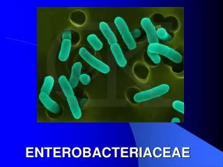
Enterobacteriaceae. Dr Ekta Chourasia Lecturer, Microbiology. General Features of Enterobacteria. Present in large intestine. Gram negative bacteria. Aerobic or facultative anaerobic. Motile by peritrichate flagella or non motile. Grow on ordinary media ( non fastidious ).

Enterobacteriaceae
E N D
Presentation Transcript
Enterobacteriaceae Dr Ekta Chourasia Lecturer, Microbiology
General Features of Enterobacteria Present in large intestine Gram negative bacteria Aerobic or facultative anaerobic Motile by peritrichate flagella or non motile Grow on ordinary media (non fastidious) Ferments glucose with acid & gas or only acid Reduce nitrates to nitrites Catalase + ve & oxidase -ve Dr Ekta Page 2
Classification of Enterobacteriaceae • Based on lactose fermentation – oldest method : • Lactose fermenters e.g. Escherichia, Klebsiella. • Late lactose fermenters e.g. Shigella sonnei • Non lactose fermenters e.g Salmonella, Shigella • Commensal intestinal bacteria: LF • Intestinal pathogens: NLF Dr Ekta Page 3
Classification of Enterobacteriaceae • Modern taxonomy – group together bacteria that possess: • Common morphological and biochemical properties • Similar DNA base compositions • Family – Tribe / Group - Genera Dr Ekta Page 4
Enterobacteriaceae(Tribes & Genera) CDC 1989 Tribe 1 Eshcherichieae Tribe 5 Klebsielleae Klebsiella Enterobacter Serratia Escherichia Hafnia Pantoea Shigella Proteus Tribe 2 Edwardsielleae Tribe 6 Proteeae Morganella Providentia Edwardsiella Tribe 7 Yersinieae Tribe 3 Salmonelleae Yersinia Salmonella Tribe 8 Erwinieae Tribe 4 Citrobactereae Erwinia Citrobacter Dr Ekta Page 5
Escherichia coli • Named after Escherich, first to describe colon bacillus • Normal flora of the human & animal intestine. • Remains viable in the feces for few days. • Detection of E.coli in the drinking water – indicates recent pollution with human or animal feces. Dr Ekta Page 6
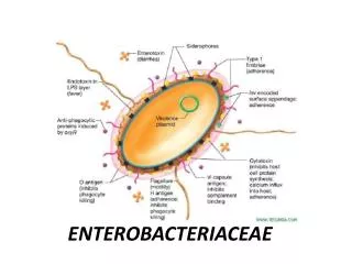
Antigenic Structure of Gram –ve Bacteria • Three antigens – serotyping of E.coli • H – flagellar antigen • O – somatic antigen • K – capsular antigen Majority do not possess K Ag. Dr Ekta Page 7
Virulence Factors - Two types of virulence factors: Surface Ags & Toxins • Surface Antigens • LPS surface O Ag – endotoxic activity, protects from phagocytosis and bactericidal effects of complement • Envelope or K Ag – protects against phagocytosis and antibacterial factors inserum • Fimbriae – colonisation factors, found in strains causing diarrhoea and urinary tract infections Dr Ekta Page 8
Virulence Factors 2. Toxins (Exotoxins) – two types • Enterotoxins – pathogenesis of diarrhoea - 3 types : LT (heat labile toxin), ST (heat stable toxin) & VT (verocytotoxin or shiga- like toxin) • Hemolysins – may be nephrotoxic Dr Ekta Page 9
Heat Labile Toxin (LT) • Resembles cholera toxin in its structure, function and mode of action • Complex of polypeptide subunits. • LT: one subunit of A (action- enzymic), five subunits of B (binding) Dr Ekta Page 10
Heat Labile Toxin (LT) Escherichia coli / Vibrio cholerae Gut lumen Intestinal epithelial cell Dr Ekta Page 11
Heat Labile Toxin (LT) Activates Adenyl cyclase increased production of cAMP Increased secretion of Na, Cl and water from the cell Heat Stable Toxin (ST) Activates guanyl cyclase Increased production of cGMP Inhibition of ionic uptake in intestinal cells Osmotic loss of water from cells E.coli toxins Dr Ekta Page 12
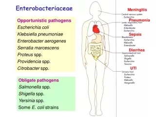
Pathogenicities/ Clinical Infections • Urinary tract infection • Diarrhoea • Pyogenic infections • Wound infection, especially after surgery of lower intestinal tract. • Peritonitis. • Biliary tract infection. • Neonatal meningitis. • Septicemia – can lead to fatal conditions like • Septic shock • Systemic Inflammatory Response Syndrome Dr Ekta Page 13
Lab Diagnosis of UTI Specimens Urine Mid stream urine (MSU) Catheter specimen urine (CSU) Supra pubic aspiration (SPA) Microscopy Wet mount Pus cells / hpf Bacteria / crystals/ casts Gram stain Gram negative bacteria (1bacteria / oil field is significant) Urine Culture Kass semi-qauntative method Standard loop technique To know significant bacteriuria Dr Ekta Page 14
Lab Diagnosis of E. coli UTI Significant bacteriuria > 105 organism / ml of MSU Culture BA / MAC : LF (flat) Identification tests I M Vi C test: + + - - TSI agar Acid, no gas AST Dr Ekta Page 15
Diarrheagenic E.coli • Enteropathogenic E.coli (EPEC) • Enterotoxigenic E.coli (ETEC) • Enteroinvasive E.coli (EIEC) • Enterohemorrhagic E.coli (EHEC) or Verotoxigenic E.coli (VTEC) • Enteroaggregative E.coli (EAEC) : “stacked brick” appearance. • Diffusely adherent E.coli (DAEC) Dr Ekta Page 16
Enteropathogenic E.coli (EPEC) • Infantile diarrhea • Institutional outbreaks • Noninvasive, nontoxigenic • Pathogenesis – adhesion via fimbria, disruption of brush border microvilli • Clinical features – fever, diarrhea, vomiting, nausea, non bloody stools • Lab Diagnosis – testing colonies grown on BA/ MA with EPEC O antisera Dr Ekta Page 17
Enterotoxigenic E.coli (ETEC) • Traveller’s diarrhea • Resembles cholera • Noninvasive, toxigenic • Pathogenesis – production of plasmid coded toxins(LT/ ST) • Clinical features - Diarrhea, vomiting and abdominal pain • Lab Diagnosis – demonstration of enterotoxin by in vitro or in vivo methods, detection of LT/ St by gene probes Dr Ekta Page 18
Enteroinvasive E.coli (EIEC) • Bloody diarrhea (dysentery), resembles Shigella dysentery • Passage of blood, mucus & leucocytes in stool • Pathogenesis - Invades epithelial cells by endocytosis and can spread laterally to adjacent cells, causes tissue destruction, necrosis and ulceration. • Lab Diagnosis: • Sereny test- instillation of suspension of freshly isolated EIEC or Shigella in the eyes of guinea pig – mucopurulent conjunctivitis and severe keratitis • Penetration of HeLa or Hep2 cells in tissue culture Dr Ekta Page 19
Enterohemorrhagic E.coli (EHEC) • Produces verocytotoxin (VT), a shiga-like toxin (SLT); hence also known as Verocytotoxigenic E.coli (VTEC) • Pathogenesis – EHEC attaches to the colonic mucosa and releases VT. VT targets vascular endothelial cells, inhibits protein synthesis - cytotoxicity • Clinical features - Mild diarrhea (bloody) to fatal complications (esp. in young children and elderly): • Hemorrhagic colitis – destruction of mucosa followed by hemorrhage. • Hemolytic Uremic syndrome – triad of acute renal failure, hemolytic anemia and thrombocytopenia. • Serotype O157: H7 is most commonly involved. • Outbreaks of food poisonings (fast foods, contaminated hamburgers) Dr Ekta Page 21
Enterohemorrhagic E.coli (EHEC) • Lab Diagnosis: • Demonstration of bacilli or VT in feces or in culture • Sorbitol MacConkey agar for O157:H7 – does not ferment sorbitol unlike other E.coli • Cytotoxic effects on Vero or HeLa cells • DNA probes to detect toxins Dr Ekta Page 22
Enteroaggregative E.coli (EAEC) • Persistent diarrhea in children in developing countries. • Aggregate to give a “Stacked brick appearance” on Hep2 cells or glass (due to fimbria) • Pathogenesis – shortening of villi, mucus biofilm, heat stable cytotoxin (hemorrhagic necrosis and edema) Dr Ekta Page 23
Epidemiology & Treatment Epidemiology • EPEC & ETEC - most important causes of diarrhea globally • EHEC – in developed countries. Treatment • Based on symptoms: • Primary treatment – fluid replacement • Secondary treatment – antibiotics in severe cases with systemic involvement Dr Ekta Page 24
Klebsiella Normal gut flora in the intestine Gram negative coccobacilli (short & plump) Capsulated, non-motile, Mucoid LF colonies on MAC Species Pneumonia, Urinary tract infections K. pneumoniae K. oxytoca Atrophic rhinits K. ozaenae Rhinoscleroma K. rhinoscleromatis Dr Ekta Page 25
Pathogenicities of Klebsiella pneumoniae • Pulmonary infections - Pneumonia (lobar): • High fatality • In middle aged or older persons with medical problems like DM, alcoholism, chronic bronchopulmonary disease • Extensive necrosis & hemorrhage resulting in thick, mucoid, brick red sputum “currant jelly like” • Extrapulmonary infections – • Meningitis & enteritis in infants • UTI • Septicemia • An important cause of nosocomial infections. Dr Ekta Page 26
Lab Diagnosis - Klebsiella Specimens Urine, sputum, nasal secretions / swab, blood Culture BA / MAC : LF (mucoid) Identification tests I M Vi C test: - - + + TSI agar Acid with gas Urease Positive Dr Ekta Page 27
Proteus Normal gut flora in the intestine Gram negative bacilli, pleomorphic Motile, Non lactose fermenter NLF on MAC Swarms on BA, Urease +, H2S + Species P mirabilis P vulgaris UTI Pneumonia Wound infections Urease converts urea to NH4 & CO2 causing alkalinization of urine leading to renal calculi (stones) Proteus antigens are used in the Weil - Felix test to diagnose Rickettsial diseases Dr Ekta Page 28
Lab Diagnosis - Proteus Specimens Urine, sputum, wound swab BA: swarming Culture MAC : NLF, fishy/ seminal smell Identification tests Urease + Indole: PM - / PV + TSI agar K / A (H2S) Dr Ekta Page 29
Enterobacter, Serratia, Citrobacter • Moist environments in hospitals – common reservoirs. • Pathogenicities – • UTI, • Wound & respiratory infections in hospitalized patients, • Outbreaks in ICUs, burn units & other special units Dr Ekta Page 30