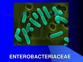
Enterobacteriaceae
370 likes | 585 Vues
Enterobacteriaceae. Indications for stool culture include. Bloody diarrhea Fever Tenesmus (is the constant feeling of the need to empty the bowel, accompanied by pain, and cramping) Severe or persistent symptoms Recent travel to a third world country Known exposure to a bacterial agent

Enterobacteriaceae
E N D
Presentation Transcript
Indications for stool culture include • Bloody diarrhea • Fever • Tenesmus (is the constant feeling of the need to empty the bowel, accompanied by pain, and cramping) • Severe or persistent symptoms • Recent travel to a third world country • Known exposure to a bacterial agent • Presence of fecal leukocytes
Aim of the test • Detect bacterial pathogenic organisms in the stool; diagnose typhoid fever, • enteric fever, bacillary dysentery, Salmonella infection
Types of specimen • Stool or rectal swab or stool (fresh random) in fecal transport system
Criteria of specimen rejection Formed stool, specimen contaminated with urine, residual soap, or disinfectants. Specimens received in grossly leaking transport containers; diapers; dry specimens; specimens submitted in fixative or additives;
In acute or subacute diarrhea, three common syndromes are recognized: gastroenteritis, enteritis, and colitis (dysenteric syndrome). With colitis, patients have fecal urgency and tenesmus. Stool are frequently small in volume and contain blood, mucus, and leukocytes. External hemorrhoids are common and painful.
Diarrhea of small bowel origin is indicated by the passage of few large volume stools. This is due to accumulation of fluid in the large bowel before passage. Leukocytes indicate colonic inflammation rather than a specific pathogen. Bacterial diarrhea may be present in the absence of fecal leukocytes and fecal leukocytes may be present in the absence of bacterial or parasitic agents (ie, idiopathic inflammatory bowel disease).
Although most bacterial diarrhea is transient (1-30 days) cases of persistent symptoms (10 months) have been reported. The etiologic agent in the reported case was Shigella flexneri diagnosed by culture of rectal swab. In infants younger than 1 year of age, a history of blood in the stool, more than 10 stools in 24 hours, and temperature greater than 39°C have a high probability of having bacterial diarrhea. Diarrhea is also a common side effect of long term antibiotic treatment. Although often associated with Clostridium difficile, other bacteria and yeasts have been implicated.
Specimen collection • A single stool specimen cannot be used to rule out bacteria as a cause of diarrhea. More than two specimens should only be submitted from patients for whom there is a high degree of suspicion. The stool should be collected on collected in sterile bedpan. A sample is transferred with the sticks to the container. The specimen should contain at least 5 g of faeces and, if present, those parts that contain blood and/or mucus should be selected. The specimen should not be contaminated with urine. Close the lid.
Rectal swab • Pass swab beyond anal sphincter, carefully rotate, and withdraw. Swabbing of lesions of rectal wall or sigmoid colon during proctoscopy or sigmoidoscopy is preferred. • Duodenal or sigmoid aspirate: Specimen should be collected by a physician trained in this procedure
Time relapse before processing the sample • Stool samples should be examined and cultured as soon as possible after collection. As the stool specimen cools, the drop in pH will inhibit the growth of most Shigella spp. and some Salmonella spp. • Storage: Refrigerated (2-8 °C)
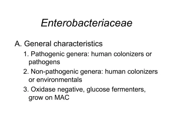
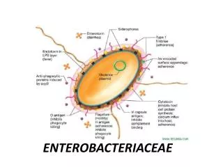
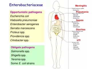
E.Coli (Coli form group) • Morphology: G-ve Bacilli, Motile, Non-spore , Capsulated in pathogenic strains. • Media: Ordinary, MacConkey (Pink colony) Facultative anaerobic, Co2. Temp:37C
Types of E.coli • Enteropathogenic: diarrhea via adhesion of M.O to epithelial cells • Enteroinvasive: diarrhea via invasion of intestinal epithelium • Enteroaggregative: diarrhea+colitis • Enterotoxogenic: diarrhea by exotoxin • Enterohemorrhagic: diarrhea via verotoxin (haemorrhagic coltis)
Other types of E.coli • Urinary tract infection ( Urine) • Meningitis (C.S.F) • Pneumonia(Sputum) • Endocarditis ( • Septicemia (Blood) • Wound infection (pus) • Appendicitis (blood). (protenial fluid) • Cholycystitis (urine) • Diarrhea (stool)
Limitations • Yersinia sp, Vibrio, E. coli O157:H7, and Campylobacter spp. will not be isolated unless specifically requested; These organisms are fastidious and have very specific requirements for growth.
Stool Culture, E. coli O157:H7 • Aim of the test: Detect E. coli O157:H7 from stool specimen or rectal swab and perform sensitivity test. The Latex test wil demonstrate by slide agglutination, E. coli strains possessing the somatic O157 antigen and Flagellar H7 antigen.
Media :Sorbitol MacConkey Agar (SMAC) • A loopful of stool is streaked on Sorbitol MacConkey. Incubate at 37 oC. Under aerobic conditions. Examine plates for non-sorbitol fermenting colonies (NSF). • NSF colonies may be taken from SMAC plates or alternatively NSF isolates may be inoculated onto non-selective agar media for testing. It is necessary to test up to 10 separate NSF colonies to ensure a high probability of detection from mixed cultures.
LATEX PROCEDURES • Bring the latex reagents to room temperature. Make sure the latex suspensions are mixed by vigorous shaking. Expel any latex from the dropper pipette for complete mixing. • Dispense 1 drop of the Test latex onto a circle of the black slide. Place it close to the edge of the circle.. • Add some loopfuls or a Pasteur pipette drop of saline to the circle. Ensure that the latex and saline do not mix at this stage. • Using a loop, pick off a portion of the colony to be tested and carefully emulsify in the saline drop.
5. Mix the Test latex and suspension together and spread to cover most of the reaction area using the loop. Flame the loop. 6. Rock the slide in a circular motion, observing for agglutination. Do not rock the card for more than 1 minute and do not use a magnifying glass. 7. If no agglutination occurs, then proceed to test other NSF colonies if these are present. 8. If agglutination with the test reagent does occur, then it is necessary to test a further portion of the colony with the control reagent to ensure that the isolate is not an autoagglutinating strain. 9. When finished, dispose of the reaction slide into disinfectant.
Stool Culture, Vibrio spp. • Media: • Alkaline peptone water • TCBS (Thiosulfate Citrate Bile salt Sucrose Agar) • Culturing procedure: A loopful of stool is streaked onto the surface of a TCBS plate and about one gram is inoculated into a tube containing alkaline peptone water, incubate at 37oC. After 6-8 hours make a subculture from the alkaline peptone water onto the surface of a new plate of TCBS. Incubate at 37oC for 24 hours. See schematic diagram
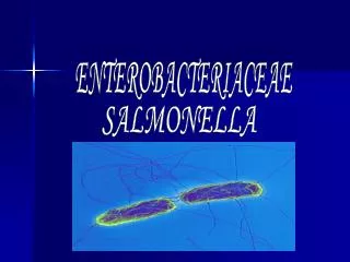
Salmonella • Classification: A. Salmonella causing enteric fever • S.typhi • S.paratyphi A • S.paratyphi B • S.paratyphi C B. Salmonealla causing food poisoning • S.Typhimurium • S.Enteritidis C. Salmonella causing septicemia • S.cholerae-suis • S.virchow
Enteric fever • Pathogenicity: Source of infection: Human case and Human carrier (Urine and stool) Mode of transmission: contamination food and water.
Pathogenesis: 1. M.O enter body via mouth multiplies in payer’s patches of small intestine via lymphatic into mesenteric Ln, spleen, liver this called incubation period which still (14 days) 2. M.O pass to blood and produced bacterimia with first appearance of symptoms and this called (First week) 3. M.O stimulate immune system to produced Ab and this called (2nd week).
3. M.O disappear from blood to internal organ, liver, gall bladder, spleen, kidney to be extracted in intestine again and M.O excreted in stool and urine this called (3rd week). 4. M.O are localized in peyer’s patches with typical pathology which include Necrosis, ulceration and proliferation of intestine in sever case.
Laboratory • Human case: • First week • sample: (stool) • Direct film stain by Gram stain: G-ve bacilli, motile. Non-spore, non-capsulated 3. culture characters: Blood culture 5-10ml of venous blood are added to 50 ml of broth containing 0.5% Na+ taurocholate and incubation at 37C/24h. And the subculture on:
MacConky’s medium pale yellow colonies • DCA (desoxycholate citrate agar) pale yellow • S-S pale yellow • XLD (xyline lysine dextrose agar) pink colonies Cloteculture: 5ml of venous blood are left to clot and then clote is added to 15 ml of bile salt broth the incubation 37C/24h. And subculture
2nd week: By Widal test 3rd week: sample (stool or urine) Stool sample riched by different type of M.O so to detected salmonella must be 5-10 ml of stool is added on (selneite broth) where kill all organismis except salmonella and shigella. Incubation 37C/24h and subculture
Human carrier • Sample: stool or urine • Direct film • Culture character: At first: M.O present in gall bladder (chola-mate) Patient give laxative diarrhea M.O in large amount in stool. Media: 5-10ml of stool+ salenite broth 37C/24h, subclture. Urine centerfugation then deposite is taken and examined