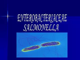
Enterobacteriaceae
Enterobacteriaceae. - Microscopic appearance. - Cultural characteristics. - Biochemical Tests of Enterobacteriaceae. - Identification of Enterobacteriaceae using API 20E. Enterobacteriaceae. - Gram negative rods. - Aerobes and facultative anaerobes. - Motile and non-motile.

Enterobacteriaceae
E N D
Presentation Transcript
Enterobacteriaceae - Microscopic appearance - Cultural characteristics - Biochemical Tests of Enterobacteriaceae - Identification of Enterobacteriaceaeusing API 20E
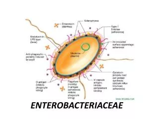
Enterobacteriaceae - Gram negative rods - Aerobes and facultative anaerobes - Motile and non-motile - Oxidase negative - Reduce nitrate to nitrite - Ferment glucose with acid production
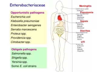
Escherichia coli - Urinary tract infections. ( urine )* - Infections of wounds, peritonitis, sepsis and toxic shocks. ( pus )* - Meningitis and bacteraemia in neonates. ( CSF & blood )* - Diarrhoeal disease. ( faeces )* *Specimens depending to site of infection.
Gram negative rod, motile. A minority of strains are capsulated.
On Blood agar E. coliproduces 1–4 mm diameter mucoid colonies. Some strains are haemolytic.
OnMacConkey agar E. coliferments lactose, producing smooth pink colonies.
On KIA (Kligler iron agar) Most strains of E. coli produce an acid deep and an acid slopewith gas production.
Klebsiellae - chect infections. ( sputum )* - Infections of wounds. ( pus )* - Urinary tract infection. ( urine )* - rhinoscleroma. ( infected tissues )* *Specimens depending to site of infection.
On Blood agar Klebsiellaeproduce large grey-white usually mucoidcolonies
On MacConkey agar Mostklebsiellae are lactose-fermenting, producing mucoidpink colonies.
Citrobacter & Enterobacter - Opportunistic pathogens occasionally isolated from urine, pus, blood and other specimens.
On MacConkey agar C.freundiiare lactose-fermenting, producing pink colonies.
On MacConkey agar Enterobacterare lactose-fermenting, producing shiny pink colonies.
Vogues Proskauer Test • used to detect acetoin in a bacterial broth culture - The test is performed by adding alpha-naphthol and potassium hydroxide to the Voges-Proskauer broth which has been inoculated with bacteria. A cherry red color indicates a positive result, while a yellow-brown color indicates a negative result
Vogues Proskauer Test - digestion of glucose to acetylmethylcarbinol. If glucose is being broken down, it will react with alpha-naphthol (VP reagent 1) and potassium hydroxide (VP reagent 2) to form a red color. Alpha-naphthol and potassium hydroxide are chemicals that detect acetoin.
Citrate Utilization Test - The test is based on the ability of an organism to use citrate as its only source of carbon. • Bacteria that can grow on this medium turn the Bromothymol • blue indicator from green to blue.
Indole Production When C.tetani is cultured in a medium which contains tryptophan. Indole production is detected by Kovac’s reagent which contains 4 (p)-dimethylaminobenzaldehyde which reacts with the indole to produce a red coloured compound.
Urease Test The test organism is cultured in a medium which contains urea and the indicator phenol red. When the strain is urease producing, the enzyme will break down the urea (by hydrolysis) to give ammonia and carbon dioxide. With the release of ammonia, the medium becomes alkaline as shown by a change in colour of the indicator to pink-red.