Download
1 / 61
871 likes | 3.07k Vues
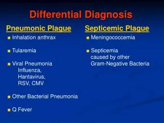
Principles of Differential diagnosis and BIOPSY. Examination and Diagnostic Methods. On detection-Present it in simple words Health history Accurate history always is helpful Oral manifestation of systemic diseases (e.g., leukemia, crohn’s disease) History of the lesion Duration

E N D
Examination and Diagnostic Methods On detection-Present it in simple words Health history Accurate history always is helpful Oral manifestation of systemic diseases (e.g., leukemia, crohn’s disease) History of the lesion Duration Change of size Change in character Symptoms – Pain, altered function, dysphagia, lymphadenopathy Associated constitutional symptoms Associated trauma
Clinical evaluation Examine in detail Regional lymphadenopathy Detailed description Draw the lesion on the chart Inspection-Describe before handling Palpation Percussion Auscultation
Clinical evaluation Anatomic location Ascertain as much as possible which tissue/s are contributing to the lesion Cause (THINK OF COMMON CAUSES FIRST) Physical character of the lesion Size and shape of the lesion Single versus multiple lesions Surface of the lesion (Smooth, lobulated, irreguar)
Clinical evaluation –Anatomic location Color Sharpness of the boundaries Consistency Fluctuation Pulsation- (vascular lesions) Lymph nodes Radiographic examination Laboratory tests
Basic Tenets of referral • Examine yourself- You make the decision • Biopsy or referral • Health of the patient • Surgical difficulty • Malignant potential
Biopsy Definition Types Oral Cytology Aspiration biopsy Incisional biopsy Excisional biopsy
Oral Cytology –based Procedures Substitutes of biopsy Types Exfoliative cytology Oral brush cytologic examination
Exfoliative Cytology Common diagnostic tool for uterine cervical cancer Results are not reliable False negative diagnoses Post operative discomfort more significant
Brush Biopsy Valuable non invasive diagnostic tool Chronic mucosal changes Good adjunct Technique is precise Cost is nominal Covered by most Insurance plans Oral brush cytologic examination Hand held rotary wire brush
Oral brush cytologic examination Local /topical anesthetic is usually not required Rotary brush is placed in contact with the surface of the lesion
Oral brush cytologic examination Brush is rotated 5-10 times with firm pressure. Epithelial cells are collected from all three layers
Oral brush cytologic examination Brush + collected material is smeared on the slide Flood with fixative material Wait to dry Categorization of specimen Negative Positive Atypical
Incisional Biopsy Small portion of lesion is removed Large lesion-More than one biopsy indicated Indications >1 cm Risky hazardous location Suspected Malignancy
Principles Wedge of tissues is excised Normal / abnormal tissue Perimeter of lesion preferred Active growth Significant cellular changes Include base of lesion Anatomic structures should be spared
Excisional Biopsy Removal of the entire lesion Smaller lesions (<1cm) Pigmented small vascular lesions Principles: Lesion + 2-3 mm of normal appearing surrounding tissue
Surgical Principles Local Anesthesia Do not inject within the tissues to be excised 4 point anesthesia technique
Tissue stabilization Movable structures Methods of stabilization Assistant Traction/Stay suture Instruments Chalazion Towel clips Non tooth tissue forceps
Hemostasis And Incision Avoid suction Incision Instruments Scalpel AVOID Electrosurgery Laser Hemostasis
Incison • Initial incision should be gauged to exceed the total depth of the lesion slightly • Elliptical incision • Two ellipse at surface • Converge like a V at the base
Thin deep specimens are preferable than broad shallow specimens Keep incisions parallel to nerves, arteries and veins
Principles Entire lesion along with 2-3mm of normal appearing tissue The margin should be increased to 5mm of normal tissue when excising pigmented, Malignant (small lesions), vascular tissues
Tissue handling Ensure tissue is NOT : Crushed For small biopsies- Use instruments with caution USE of traction suture through the specimen avoids specimen TRAUMA
Aspiration Biopsy Needle and syringe Aspiration of contents of the lesion 2 types Explore if lesion contains fluid Aspirate cells (fine needle aspiration cytology) Soft tissue masses Neck Masses
Punch Biopsy Available in various diameters Disposable Advantages Ease of technique Smaller punch no sutures required Produces acceptable specimen in bound down tissue Disadvantages May not be adequate for biopsy of deeper pathology Difficult on movable tissues
Punch Biopsy Procedure performed by Punch biopsy forceps
Biopsy Data Sheet Label Demographic data Pertinent history Clinical description Radiographs Clinical Impression Follow up (10days to 2 weeks)
Intra osseous Biopsy technique Lesion on or within the bone Any lesion that is unrelated to the dentition or does not respond to customary treatment of dentition should have tissue removed for definitive diagnosis Excisional biopsy Periapical granulomas Odontogenic Cysts (radicular Cyst) Compare the lesion to contra lateral side
Technique Mucoperiosteal flap Precautionary Aspiration 18gauge needle, 5- 10ml syringe Bur hole Inability to aspirate indicates solid mass Pulsatile vascular lesion (hemangioma or AV malformation ) Osseous Window Cortical bone is penetrated through a bur Trephine drill with irrigation
Overview Odontogenic cysts & tumors arise from the odontogenic apparatus. The odontogenic apparatus consists of: Epithelium: Remnants of dental lamina Reduced enamel epithelium Odontogenic rests Lining of odontogenic cysts Basal cell layer of oral mucosa Ectomesenchyme: Dental papilla
What is a cyst? Definition: A cyst is a pathological cavity with fluid, semi-fluid or gaseous contents, which is not created by accumulation of pus. It is frequently lined by epithelium. Typical features:a) Cysts grow slowly and expansivelyb) Form sharply-defined radiolucencies with smooth bordersc) Frequently they are found as an incidental radiographic finding.
Classification Odontogenic cysts: Inflammatory: Periapical (radicular) cyst Residual periapical (radicular) cyst Developmental: Dentigerous cyst Odontogenic keratocyst (OKC) Gingival (alveolar) cyst of the newborn Gingival cyst of the adult Lateral periodontal cyst Calcifying odontogenic (Gorlin) cyst Eruption cyst Non Odontogenic cysts that are not really cysts: Nasopalatine duct cyst, Nasolabial cyst, Dermoid cyst, Cysts without epithelial lining: Simple bone cyst Aneurysmal bone cyst
Diagnostic modalities Panoramic Radiograph, CBCT are most valuable diagnostic tools for detection Histopathology Confirms diagnosis.
Clinical Features Noticeable swelling: Initially smooth bony hard swelling with normal overlying mucosa As bone thins through resorption, cyst may show through as bluish fluctuant swelling (may be compressible in nature) Discharge into mouth Pain due to secondary infection Fluid may be aspirated Thin-walled cysts may be trans illuminated
Basic Goals Eradication of Pathological condition Functional rehabilitation Broad Classification Cysts Cystlike lesions of the jaws Benign tumors of the jaws Benign tumors of the soft tissues Malignant tumors
Cysts Surgical Management Four methods Enucleation Marsupialization A staged combination of two procedures Enucleation with curettage
Enucleation Definition : Shelling out of the entire lesion without rupture Indications Any cyst that can be removed without sacrificing any adjacent vital structures. Advantages Cyst is removed entirely Complete Histopathology Disadvantages Devitalization of teeth Jaw fracture w/o curettage OKC may be left behind