Download
1 / 62
660 likes | 931 Vues
Cell Cycle and Cell Division. Prokaryotic Cells - Bacteria. E. coli is a bacteria that resides in our colon and can be beneficial to us – our major source of vitamin K that is necessary for proper coagulation

E N D
Prokaryotic Cells - Bacteria • E. coli is a bacteria that resides in our colon and can be beneficial to us – our major source of vitamin K that is necessary for proper coagulation • Have only 1 copy of their DNA, considered to be haploid, any change in the sequence may be readily seen in the phenotype – observable characteristics
Binary Fission • E coli can grow and duplicate every 20 minutes • DNA is attached to the cell membrane and after it duplicates, it separates as the cell enlarges • When cell is double in size, the cell divides by adding a plasma membrane and cell wall between the 2 cells
Escherichia coli Genome • DNA is circular and encodes ~ 3000 proteins • Bacteria with unusual growth properties have been identified – contain mutations that changed the phenotype • Rifampicin growth because of mutation in the RNA polymerase that allows for mRNA synthesis while in presence of the drug • Bacteria can pass DNA back and forth between one another • Mixed 2 mutant types together and found that bacteria can grow on media without either amino acid
Gene Transfer • Bacterial mating or conjugation – can be achieved in bacteria that contain plasmids– small, circular dsDNA that is separate from rest of chromosome • Plasmids can replicate independently from the bacterial chromosome • F plasmid or fertility plasmid allows for mating and gene transfer
Bacterial Mating • Bacteria with F plasmid (donor) will encounter a bacteria without the F plasmid (recipient) and establish a cytoplasmic bridge to pass on the F plasmid • Now both are donor bacteria • Method of passing on antibiotic resistance
Integration • The plasmid can integrate into the chromosome and when bacteria conjugate they can pass on chromosomal genes • Increases genetic variation
Transformation • Bacteria can pick up DNA from surroundings • Take advantage of this in molecular biology labs to replicate DNA fragments of choice
DNA in the Eukaryotic Cell • A cell’s DNA is divided into a set of chromosomes – each being a long linear DNA associated with proteins to help fold into chromatin • Also associated with proteins involved in gene expression, DNA replication and DNA repair
Chromosomes • With exception of the sex chromosomes (X and Y) humans have 2 similar copies of each chromosome • one from our mother (♀) • one from our father (♂) • Called homologous chromosomes • One instance of non-homologous chromosomes are males because of their X and Y • Chromosomes are similar but not identical
Chromosomal Bands • Set of human chromosomes is called a karyotype • The staining patterns can allow geneticists to identify areas of abnormalities
Types of Chromosomes • Mitotic chromosomes are those that are visible during mitosis – condensed • Interphase chromosomes are loose and string-like - chromatin
Components of Chromosomes • 3 important DNA sequences needed for accurate DNA copying, complete genome to new daughter cells • DNA replication origin – a place to start DNA replication, multiple sites • Centromere – a place to attach the mitotic spindle for chromosome separation • Telomere – at end of linear DNA to prevent the chromosome from getting shorter in every round of replication
Telomeres • Telomerase is the enzyme responsible for adding DNA sequence to the end of the chromosome to act as a template to allow complete DNA replication • Also protects the DNA from degradation
Chromatin vs Chromosome • Chromatin is a complex of protein and DNA – very dispersed • Chromosome is the compact form of DNA so that it is protected during mitosis
50 nm Nucleosomes • Nucleosome – “bead-on-a-string” look to DNA • The string is the DNA and the bead is the nucleosome core particle that the DNA is wrapped around – the protein is histone
Histone Octmer • Nucleosome is the histone proteins and the adjacent linker DNA – threads the histones together • Histone is core around which the DNA coils • Disc shaped with 2 copies of H2A, H2B, H3 and H4
Histones • Proteins with a large proportion of positively charged amino acids (Lys and Arg) • Helps to bind the negatively charged DNA • 5 types in 2 main groups • Nucleosomal histones • Small and coil DNA into nucleosomes • H2A, H2B, H3 and H4 • H3 and H4 highly conserved • H1 histones • Larger and less conserved
Interphase Chromosomes • While not as condensed as mitotic chromosomes, there are areas more tightly packed than others • Depends on the genes that are being expressed • Most highly condensed chromatin is called heterochromatin • The more extended chromatin is called euchromatin and is either being transcribed or easily available for transcription
X Chromosome • Females have 2 X chromosomes and males have 1 X and 1 Y chromosome • One X becomes inactive and highly condensed – Barr body • Some cells have the paternal X off and some have the maternal X off • This happens during early development and once off, all cells that arise from that cell will have it off • This is then passed down (inherited) in all the cells that arise from that cell
Cytoskeleton’s Role • Mitotic spindle – microtubules used to separate the chromosomes – formation starts in late G2 • Contractile ring – myosin and actin filaments used to separate the 2 daughter cells – formation starts in M phase
Organelles Fragment • Chromosomes divide as discussed in Chapter 6 • Mitochondria and chloroplasts will divide and double their numbers • Membrane bound organelles fragment to increase the likelihood to be divided evenly between 2 daughter cells
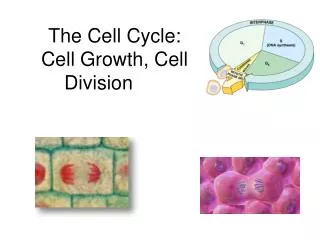
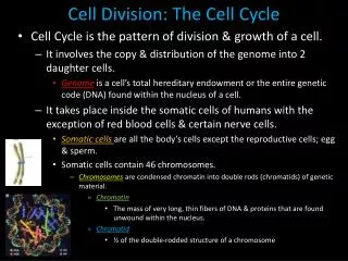
Cell Cycle • The orderly sequence of events which a cell duplicates its contents (DNA and organelles) and then divides in 2 • Fundamental task is to copy and pass on its genetic information to daughter cells
Cell Cycle • New cells need to get a copy of the entire genome in the process – most cells double their mass as well
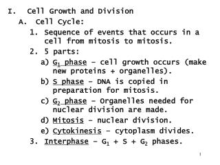
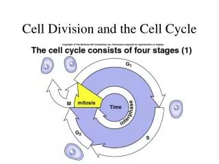
4 Phases of the Cell Cycle • Mitosis or M phase has 2 steps • Nucleus divides – mitosis • Cell divides in 2 - cytokinesis
4 Phases of the Cell Cycle • Period between mitosis is called interphase – has 3 phases • Synthesis (S) phase is when DNA is replicated • Gap1 (G1) phase is phase between M and S phase • Gap2 (G2) phase is phase between S and M phase
G1 and G2 Phases • Portion of the cell cycle that allows for the synthesis of proteins, lipids and other molecules needed for cell division • Duplicates the organelles and keeps the cells from getting smaller on each division • At end of G2, the DNA condenses into chromosomes from its normal chromatin structure
5 Stages of Mitosis • Continuous event but divided into 5 stages • Prophase – chromosomes condenses and mitotic spindle forms outside the nucleus • Prometaphase – nuclear membrane breaks down, attach mitotic spindle to chromosomes • Metaphase – mitotic spindle lines up chromosomes along the equator of the cell • Anaphase – sister chromatids separate and move to opposite ends • Telophase – nuclear envelope reassembles and cell is ready for cytokinesis
Prophase • Centrosome is duplicated in late S phase and as prophase starts they separate and move to opposite poles of the cell using motor proteins and ATP hydrolysis • Each centrosome organizes its microtubules which then interact to form the mitotic spindle which undergoes dynamic instability
Spindle Poles • During prophase some of the microtubules become stabilized and form the mitotic spindle • Some microtubules interact and are called polar microtubules and the centrosome is called the spindle pole
Prometaphase • Starts with the break up of the nuclear envelope because of the disassembly of the intermediate filaments in the nuclear lamina • Spindle microtubules will bind to the chromosomes at a structure called the kinetochore which form on chromosomes during late prophase on the centromere
Kinetochore • Kinetochore protein assembles on centromere • Microtubules probe into the area and when encounters a kinetochore will attach – now called the kinetochore microtubule • A microtubule from each pole will bind to the chromosome • Humans bind 20 to 40 microtubules per kinetochore
Metaphase • Chromosomes line up at the metaphase plate that is halfway between spindle poles • Chromosomes are constantly under tension from both kinetochore microtubules
Anaphase • The connection between chromatids is cut by proteolytic enzymes – now called daughter chromosome – pulled to the spindle pole • Process of chromosome segregation
Anaphase A and B • Anaphase A – separate the chromosomes to the spindle pole • Anaphase B – the spindle poles separate further separating the chromosomes
Telophase • Nuclear envelope reforms around each group of chromosomes • Nuclear lamina reforms • Chromosomes de-condense – return to chromatin
Cytokinesis • Separation of the new nuclei and cytoplasmic organelles between 2 new daughter cells • Starts in anaphase but is not complete until after 2 new nuclei are formed • Caused by the contractile ring of actin and myosin
Cleavage Furrow • Evidence of cytokinesis is by the presence of the cleavage furrow • Dependent on the location of the mitotic spindle • Usually forms around the equator of the cell - exception is during early development
Contractile Ring • Array of actin and myosin filaments that assembles during anaphase • Force to divide cells comes from the actin moving over the myosin getting continuously smaller • Disassembles when cell is divided into 2
Meiosis • Process of getting a cell with half the number of chromosomes – cell would be haploid • ‘Normal’ cells would have a full set of chromosomes from the mom and the dad and are called diploid • Fertilization will return the haploid sex cell to a diploid cell by uniting sperm and egg
Meiosis • Germ cells or gamates (in plants they may be spores) contain one set of chromosomes • Egg is large and non-motile • Sperm is small and motile • Diploid cells formed by fertilization
Meiosis • Involves the duplication of the chromosomes and then 2 successive cell divisions to yield the haploid cells • Can get genetic recombination between the chromosomes prior to the first cell division since they are in such close proximity – allows for new phenotypes
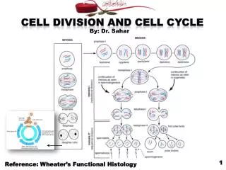
Mitosis vs Meiosis • In mitosis all 46 chromosomes line up along the metaphase plate and 1 copy of each gets taken to the new daughter cell • Each cell gets both ♀ and ♂ (46 chromosomes) • In meiosis the chromosomes find their homologous pair and these line up at the metaphase plate – get parental chromosome shuffling during the division • First division the cell gets either the ♀ or ♂ chromosome but not both (23 sister chromatids) • Second division the cell gets a single copy of the chromosome (23 chromosomes)
Difference #1 • Mitosis – chromosomes line up in the metaphase plate • Meiosis – homologous pairs line up together
Genetic Recombination • DNA can undergo rearrangements, caused by genetic recombination • General recombination – between any pair of homologous DNA sequences • 2 copies of same chromosome • Crossing over of chromosomes during meiosis • No nucleotides get altered – no gain or loss of nucleotides at the cross over point