Chromosomes, the Cell Cycle, and Cell Division
600 likes | 1.03k Vues
Chromosomes, the Cell Cycle, and Cell Division. Systems of Cell Reproduction. The continuity of life is based on the reproduction of cells. All the cells in a multicellular organism originate from a single cell (zygote). Cell division is necessary for: Reproduction Growth and

Chromosomes, the Cell Cycle, and Cell Division
E N D
Presentation Transcript
Systems of Cell Reproduction • The continuity of life is based on the reproduction of cells. • All the cells in a multicellular organism originate from a single cell (zygote). • Cell division is necessary for: • Reproduction • Growth and • Repair of an organism
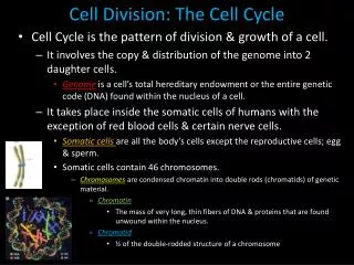
Cell Division In order for cell division to occur: • A signal to reproduce must be received. • Replication of DNA and other cell components must occur. • Distribution of replicated DNA • Cytokinesis
Prokaryotes • Have one circular chromosome. • Prokaryotes grow in size, replicate DNA and divide into 2 cells in a process called cell fission. • Initiation of fission is controlled by environmental conditions. • Cytokinesis separates the one cell into two, each with a complete chromosome.
Eukaryotes • Eukaryotic cells divide by mitosis or meiosis. • The reproduction of eukaryotic cells involves: • Replication of the DNA • Segregation of the replicated DNA into two new nuclei (nuclear division) • Division of the cytoplasm (cytokinesis) • Eukaryotes usually have many chromosomes. • Eukaryotes have a nucleus, which must replicate and, with few exceptions, divide during cell division.
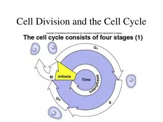
Cell Cycle • The cell cycle has two phases: mitosis and interphase. • Interphase is the period between divisions. • A typical eukaryotic cell will spend most of its life in interphase which consist of G1, S and G2. DNA is replicated during the S phase. • Most of a cell’s life is spent in interphase. • Human nerve and muscle cells, lose the capacity to divide altogether and stay in interphase indefinitely, while other cells divide regularly or occasionally.
Cell Cycle • Transitions from G1 to S and G2 to M depend upon a protein called cyclin-dependent kinase, or Cdk. • Cdk is activated by binding to a second type of protein called cyclin. • Cyclin-Cdk complexes act as checkpoints. They allow or prevent the passage to the next cell cycle stage. • In cancer cells, these cyclin-Cdk controls are often disrupted.
Figure 9.4 Cyclin-Dependent Kinases and Cyclins Trigger Transisions in the Cell Cycle
Eukaryotic Chromosomes • Chromatin is a DNA protein complex. • After DNA replication (S phase), the chromatin material condenses by wounding around histone cores. • DNA folds repeatedly becoming densely coiled and folded. • Each duplicated chromosome consists of two chromatids. They are joined at a single point called the centromere.
Mitosis: Distributing Exact Copies of Genetic Information • DNA and centrosomes duplicate in the S phase. • In animal cells each centrosome contains two centrioles. • During G2-to-M transition, the two centrosomes separate and move to opposite ends of the nuclear envelope. • Centrosomes are regions where aster rays and spindle fibers are formed.
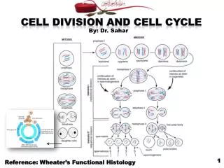
Mitosis Mitosis is divided into phases for ease of study. • Prophase • Preometaphase • Metaphase • Anaphase and • Telephase
Figure 9.8 – Part 1 figure 09-08a.jpg Figure 9.8 – Part 1
Figure 9.8 – Part 2 figure 09-08b.jpg Figure 9.8 – Part 2
Mitosis: Distributing Exact Copies of Genetic Information • Prophase marks the beginning of mitosis. • Chromosomes condense and appear as chromatids. • The chromatids are held together by the centromere. • Nucleoli disappear. • Spindle fibers form between the two centrosomes. • Radiating from the centrosome are aster rays. • Kinetochores develop.
Prometaphase • During prometaphase the nuclear envelope disappears – it breaks into small vesicles, permitting the fibers of the spindle to “invade” the nuclear region. • The chromosomes move toward the middle of the spindle. • The spindle fibers attach to the chromatids at the kinetochores.
Metaphase During metaphase, • the double chromsomes (consisting of 2 chromatids) aggregate at the equatorial plate. • the double chromosomes are readily distinguishable. Karyotypes are done using chromosomes from this phase. • the kinetochores attach to the spindle fibers. • at the end of metaphase, all chromatid pairs separate simultaneously.
Anaphase • Anaphase begins when the chromatids separate. • Motor proteins and shortening of kinetochore move a chromatid (now referred to as a chromosome) to opposite poles. • When chromosomes top moving the cell enters telophase.
Telophase • The chromosomes uncoil to form tangled chromatin. • Nuclear envelopes and nucleoli re-form forming two nuclei whose chromosomes are identical to each other and to those of the cell that began the cycle.
Cytokinesis: The Division of the Cytoplasm • Animal cells divide by a furrowing (a “pinching in” or constriction) of the plasma membrane. • In plant cells, cytokinesis is accomplished by vesicle fusion (from the Golgi apparatus) producing a cell plate.
Reproduction: Asexual and Sexual • Mitosis can repeat many times resulting in vast numbers of genetically identical cells. • Mitosis video
Reproduction: Asexual and Sexual • It is possible to stain and photograph chromosomes during metaphase. • A photograph of the slide can be taken, and images of each chromosome can be organized based on size, shape, banding patterns, and location of centomere. • This organization is called a karyotype.
Control of Cell Division • Proteins form Cdks complexes that control all stages of the cell cycle. • Cdk complexes are composed of cyclin, a protein whose concentration cyclically fuctuates, and a kinase.
Control of Cell Cycle Cyclin-Cdk complexes act as checkpoints • If DNA is damaged by UV radiation, p21 stops the cell cycle until DNA is repaired. • Cyclin-Cdk defects have been found in cancer cells. • A breast cancer with too much cyclin D has been found. • Protein 53, which inhibits activation of Cdk is found defective in ½ of all human cancers.
Control of Cell Division • External controls also influence the cell cycle. • Growth factors – a growth factor is a protein that stimulates other cells to divid. • Density-dependent inhibition. • Anchorage dependence.
Reproduction: Asexual and Sexual • Asexual reproduction produces a new organism genetically identical to the parent. • Any genetic variety is due to mutations.
Sexual Reproduction • Two gametes each containing ½ of the number of chromsomes found in other cells combine • A single cell is formed called the zygote, or fertilized egg. • Fosters genetic diversity. • Meiosis is involved.
Genetic Diversity • In each pair of chromosomes one chromosome comes from the mother and the other from the father. • The members of the pair are called homologous chromosomes The chromosomes are similar in size and appearance but different in DNA composition. • Haploid cells, 1n, contain 1 homolog from each pair. Example – gametes • When haploid gametes fuse in fertilization, they create the zygote, which is 2n, or diploid.
Meiosis: A Pair of Nuclear Divisions • Meiosis consists of two nuclear divisions that reduce the number of chromosomes to the haploid number. The DNA is replicated only once. • The functions of meiosis are: • To reduce the chromosome number from diploid to haploid. • To ensure each gamete gets a complete set of chromosomes. • To promote genetic diversity among products.
Figure 9.14 – Part 1 figure 09-14a.jpg Figure 9.14 – Part 1
Figure 9.14 – Part 2 figure 09-14b.jpg Figure 9.14 – Part 2
Meiosis: A Pair of Nuclear Divisions • Meiosis I is preceded by an interphase in which DNA is replicated. • During prophase I, synapsis occurs: The two homologs are joined together by a complex of proteins. • Genetic material may be exchanged between homologous chromosomes - crossing-over. • This crossing-over increases genetic variation by reshuffling the genes on the homologs.
Figure 9.16 Crossing Over Forms Genetically Diverse Chromosomes
Meiosis: A Pair of Nuclear Divisions • In metaphase I, the paired homologs gather at the equator. • The homologous chromosomes separate in anaphase I. • The individual chromosomes are pulled to the poles, with one homolog of a pair going to one pole and the other homolog going to the opposite pole. • Result is two nuclei each with the haploid number of chromosomes with two sister chromatids.
Meiosis: A Pair of Nuclear Divisions • The second meiotic division separates the chromatids. • Meiosis II is similar to mitosis however DNA does not replicate before meiosis II. • In meiosis II, there is no crossing-over. • Result is 4 cells each with a haploid number of chromosomes.
Meiosis: A Pair of Nuclear Divisions • Meiosis leads to genetic diversity. • Synapsis and crossing-over during prophase I mix the genetic material. • Which member of a homologous pair segregates or goes to which daughter cell at anaphase I is a matter of chance. This phenomenon is called independent assortment.
Nondisjunction • Nondisjunction can occur when homologous chromosomes fail to separate during anaphase I, or. • if sister chromatids fail to separate during anaphase II