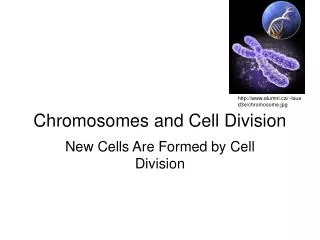
Chromosomes and Cell Division
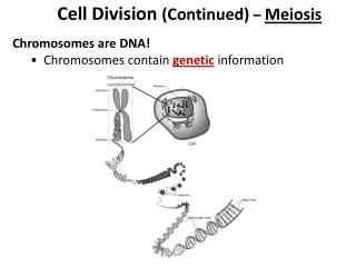
Chromosomes and Cell Division. Chromosomes. Visible only during cell division. Most condensed form of DNA. DNA double helix tightly coiled around proteins. After DNA replication. Prior to DNA replication. Chromatin = loosely coiled form of DNA An interphase cell has no visible chromosomes.

Chromosomes and Cell Division
E N D
Presentation Transcript
Chromosomes • Visible only during cell division. • Most condensed form of DNA. • DNA double helix tightly coiled around proteins.
After DNA replication Prior to DNA replication Chromatin = loosely coiled form of DNA An interphase cell has no visible chromosomes. Although not visible, every chromosome in a non-dividing cell is made up of only 1 chromatid. Only if a cell is preparing to divide, does a chromosome become made up of 2 chromatids.
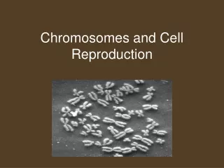
Human Chromosomes 46 Chromosomes 23 pairs
Diploid Cells • Diploid = 2n • n = # of types (sizes) of chromosomes • n = 23 for humans, so the diploid # for humans is 46 (2 x 23) • Diploid cells have pairs of chromosomes • Homologous chromosomes: chromosomes of the same shape and size, carry genes for the same traits. • Each species has a unique diploid number.
Homologous Chromosomes e.g.: eye color From “Dad” From “Mom”
Fertilization produces diploid cells • Example: Humans • The mother’s egg cell has 23 chromosomes • The father’s sperm cell has 23 chromosomes • When these cells fuse, the fertilized egg has 46 chromosomes (23 pairs); it is diploid. • Mitosis produces trillions of body cells that are all diploid. • Each of the 23 pairs of chromosomes is called a “homologous pair”.
Haploid cells • Gametes (reproductive cells) • Egg cells (ova) and sperm cells • Produced in the reproductive organs, ovaries or testes • Haploid = 1n • n = 23 for humans so human haploid cells contain 23 chromosomes. • Haploid cellshave one of each chromosome; they do NOT have pairs of chromosomes.
Diploid vs Haploid Cells Only 1 of each type of chromosome is present in the cell. 2 of each type of chromosome is present in the cell. Note: the chromosomes in both pictures have 1 chromatid each. The # of chromatids does NOT relate to diploid/haploid.
Cells must be haploid to maintain the chromosome # of a species during reproduction • If reproductive cells were diploid, then after fertilization a human zygote (fertilized egg) would have 96 chromosomes. • To have the 46 chromosomes of a typical human cell, each reproductive cell should only have 23 chromosomes.
Meiosis produces Haploid cells • Involves 2 cell divisions: Meiosis I and Meiosis II. • Each cell division consists of PMAT. • Results in 4 cells.
For cell division, what needs to occur within a cell? • 2 copies of every DNA molecule • DNA replication during the S phase of interphase • DNA packaged into easily moveable units • Prophase: DNA “condenses” (becomes more tightly coiled around proteins) • Dissolving of the nuclear membrane to allow for distribution of genetic material (prophase) • Structures to attach to, and physically move, the chromosomes • Spindle fibers (microtubules) • Made by centrosomes
An organized way of distributing the genetic material equally between two cells. • Metaphase and Anaphase • Separation of the material into distinct cells. • Telophase: formation of new nuclear membranes, cytokinesis (division of cytoplasm)
Meiosis I • Prior to the beginning of cell division, DNA is copied (replicated). • Prophase I: • Similar to mitosis: dissolving of nuclear membrane, condensing of chromosomes, formation of spindle fibers. What is unique is: • Synapsis: pairing of homologous chromosomes (formation of tetrads). • Crossing-over: chromatids of homologous chromosomes exchange genetic material. • Results in genetic recombination: a new, unique combination of genes.
Prophase I • Letter T = tetrads • Each red box surrounds a tetrad. • Letter C = centrosomes • Produce spindle fibers. • Crossing-over is occurring in the lower tetrad.
Metaphase I • Lining up of tetrads (homologous pairs) along the “equator” of the cell. • The arrangement of the chromosomes is random. • This allows for “independent assortment”. • The random distribution of genes from different chromosomes to different gametes. • Genetic recombination: new, unique combination of genes will be present in the daughter cells.
Metaphase I • Chromosomes arranging like in “A” is just as likely as chromosomes arranging like in “B”. • Each time cells in the testes/ovaries go through meiosis, unique cells are produced.
How many arrangements are possible? • For our simulation? • For humans?
How many arrangements are possible? • For our simulation: • 4 possibilities • 2n = 23 = 8 • For humans: • 2n = 223 = 8,388,608 • Wow! • And this doesn’t even include recombination resulting from crossing-over!
Anaphase I • Separation of homologous chromosomes to opposite sides of the cell. • Sister chromatids do NOT separate. • The direction they are pulled depends on their alignment during metaphase I. • Chromosomes are “independently assorted”.
Telophase I: • Chromosomes are segregated at opposite poles, one group on each side of the cell. Cytokinesis begins, forming two daughter cells. Each chromosome consists of two chromatids.
Telophase I: • Are the new cells diploid or haploid? • HAPLOID • Only one of each chromosome
Meiosis II • After PMAT I the daughter cells are haploid. So why round two of PMAT? • The chromosomes are still in a “duplicated” state, two DNA molecules per chromosome. • The normal state of genetic material in a cell is one DNA molecule per chromosome.
Prophase II Nucleus dissolves and spindle fibers form again
Metaphase II Chromosomes line up “single-file” along the midline of the cell.
Anaphase II Sister chromatids of each chromosome detach from each other and move to opposite sides of the cell.
Telophase II A nuclear membrane forms around each set of chromosomes and cytokinesis begins, forming 4 daughter cells.
After Telophase II and Cytokinesis • A new nuclear membrane should be shown. • Cells are in Interphase.