The Aortic Arches
The Aortic Arches. Objectives. Describe the formation of the aortic arches. Enlist the derivatives of aortic arches. Discuss the development of venous system of the heart. Differentiate between fetal and neonatal circulation. Discuss the congenital anomalies of the aortic arches.


The Aortic Arches
E N D
Presentation Transcript
Objectives • Describe the formation of the aortic arches. • Enlist the derivatives of aortic arches. • Discuss the development of venous system of the heart. • Differentiate between fetal and neonatal circulation. • Discuss the congenital anomalies of the aortic arches.
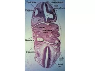
Aortic Arches • The aortic arches are a series of paired arterial channels encircling the embryonic pharynx • They: • Develop in the 4th week • Supply the developing pharyngeal arches • Arise from the aortic sac • Run dorsally, embedded in the mesenchyme of the pharyngeal arches and • Terminate in the right and leftdorsal aortae
Develop in a craniocaudal sequence There are potentially six pairs, but the fifth pair is poorly developed and disappears soon after formation Not all the 6 pairs present at the same time. By the time the 6th aortic arches are formed, the 1st & 2nd have disappeared In the region of aortic arches, the dorsal aortae remain paired, but caudal to this region they fuse to form a single median vessel Aortic --sac
During week 6 to 8, the primitive aortic arch pattern is transformed into the adult arterial arrangement of carotid, subclavian, and pulmonary arteries
First Pair • Largely disappear • Dorsal part persists as the maxillary arteries which supply the ear, teeth and muscles of the eyes and face • May give rise to the external carotid artery The first arch is obliterated before the 6th arch is formed
Second Pair • Largely disappear • Dorsal part persists as the hyoid and stapedial arteries
Third Pair • Proximal part: forms the common carotid arteries • Distal part: joins the dorsal aortae to form the internal carotid arteries
Fifth Pair • Disappears completely with NO vascular derivatives
The fate of 4 & 6thpairs of aortic arches differs on the right and left side
Fourth Pair • RIGHT: Becomes the proximal part of the right subclavian artery • LEFT: Forms part of the arch of aorta
Arch of Aorta Derived as: • Proximal segmentfrom aortic sac • Middle segmentfrom the left 4th aortic arch • Distal segmentfrom the left dorsal aorta
Subclavian Artery • The rightsubclavian artery formed from the: • Right 4th aortic arch • Right dorsal aorta & • Right 7thintersegmental artery • The leftsubclavian artery formed from the left 7thintersegmental artery
Sixth Pair • RIGHT: • Proximal part: persists as the proximal part of the right pulmonary artery • Distal part: degenerates • LEFT: • Proximal part: persists as the proximal part of the left pulmonary artery • Distal part: forms ductusarteriosus, a shunt between pulmonary artery and dorsal aorta
Changes in the original aortic arch system • Obliteration of: • Most of the 1st & 2nd arches • 5th arches completely • Distal part of the right sixth arch • The segment of both aortaelying between the 3rd & 4th arches • The segment of right aorta lying between the 7thintersegmental artery & the fused dorsal aortae
Coarctation of Aorta • Characterized by narrowing of aorta • More common in males • Classified as Preductal & Postductal types, but mostly the constriction lies distal to the origin of subclavian artery opposite the ductus arteriosus (Juxtaductal) • Preductal type: • Less common. • The narrowing is proximal to the ductus arteriosus. • If severe, blood flow to the aorta distal to the narrowing (supplying lower body) depends on a patent ductus arteriosus, and hence its closure can be life-threatening.
Postductal type Most common. The narrowing is distal to the ductus arteriosus. The ductus usually remains open to communicate pulmonary artery with the descending aorta Even with an open ductus arteriosus blood flow to the lower body can be impaired. Allows development of collateral circulation during the fetal period. The collateral circulation will develop mainly by branches from both subdavian arteries, scapular, internal thoracic and intercostal arteries. It is associated with notching of the ribs, hypertension in the upper extremities, and weak pulses in the lower extremities.
Right Arch of Aorta Occurs when theentire right aortic arch persists&thesegment of left dorsal aorta distal to the 7th intersegmental artery involutes • TYPES: • Without retropharyngeal component: The DA passes from right pulmonary artery to right arch of aorta. No effect on the trachea & esophagus • With retropharyngeal component: The right arch lies posterior to esophagus. The attachment of DA to distal part of the arch of aorta forms a ring around the trachea & esophagus and may lead to their compression
Double Arch of Aorta • Characterized by a vascular ring encircling the trachea and esophagus, usually causing compression of both structures. • The degree of compression varies • Usually the right arch is larger and passes posterior to the esophagus • The right common carotid and subclavian arteries arise separately from right arch RCC LCC RSA LSA
Patent DuctusArteriosus • Before birth, the aorta and the pulmonary artery are normally connected by a blood vessel called the ductus arteriosus, which is an essential part of the fetal circulation. • After birth, the vessel is supposed to close within a few days. The obliterated vessel forms the ligamentum arteriosum.
In some babies, the ductus arteriosus remains open (patent). This allows blood to flow directly from the aorta into the pulmonary artery, which can put a strain on the heart and increase pressure in the pulmonary circulation
Abnormal Right Subclavian Artery • May arise from the distal part of arch of aorta • In some cases, the right subclavian artery arises from the descending aorta and runs behind the trachea and the esophagus to supply the right upper limb
Thank You & Good Luck