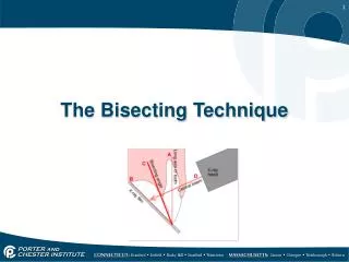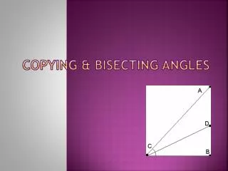
Bisecting angle technique
Bisecting angle technique. patient holds the film or sensor with his finger short cone magnified apexes. Parallel technique. film/sensor holder long cone. Image quality. small focal spot 0.7mm small unsharpness optional long cone small unsharpness

Bisecting angle technique
E N D
Presentation Transcript
Bisecting angle technique • patient holds the film or sensor with his finger • short cone • magnified apexes
Parallel technique • film/sensor holder • long cone
Image quality • small focal spot 0.7mm • small unsharpness • optional long cone • small unsharpness • geometric requirements for good images are met
Image quality • High-voltage generator • constant potential (DC) • X-ray generation • high operating frequency (66 kHz) • 25% less radiation to patient compared to conventional AC generators • shorter exposure times • improved contrast • is not affected by line voltage variations • readiness for digital systems
Kilovolts and image quality lower anode voltage higher contrast, more suitable for endodontic, apex and bone structure diagnosis medium anode voltages boarder grey scale, suitable for caries detection higher anode voltages longest grey scale spectrum for periodontal disease diagnosis 50 kV 60 kV 70 kV
Milliampers and image quality • with variable milliamperes (2 - 8 mA) we can take the whole advantage of the modern digital imaging systems and new high-speed films
Adjustable settings • adjustable kV setting (50, 53, 55, 57, 60 ,63, 66, 70) • different diagnostic needs are fulfilled • adjustable mA (2-8 mA) • maximum dose reduction possible with modern digital sensor systems and high-speed films
X-ray arm • smooth movements • drift-free positioning • no vibrations • easy, quick and accurate positioning
X-ray tube design • non-symmetric form • tubehead and cone have a common smooth plane • easy targeting along the smooth surface • close to the patient’s chest in occlusal images
Controls • 66 preprogramed quick settings • modality selection to choose film, imaging plate or sensor • density setting • adult/child selection • periapicals for different teeth • Occlusal • bite-wing / endo Quick setting allow ALWAYS to have right exposure values for individual cases
Control panel options • hand held control panel • remote exposure station • all controls easily at hand for exposure
Standard wall-mount • 4 extension arm lengths • reach 1525 – 1975 mm • special lengths available with custom order up to 2300 mm
Other mounting alternatives • dental unit mount • mobile base mount • ceiling mount • ceiling mount with operating light • floor column mount • single stud mount • pass-through mount
Imaging techniques The smaller the X-ray tube focus the smaller unsharpness small focus large focus small unsharpness large unsharpness
Imaging techniques The shorter the object-film distance the smaller the geometric unsharpness long short small unsharpness large unsharpness
Imaging techniques Geometric unsharpness and magnification decrease with a long cone long cone short cone unsharpness small unsharpness less magnification