Download
1 / 57
640 likes | 1.86k Vues
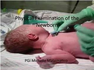
Physical examination of Newborn By Dr behzad barekatain MD Assistant professor of pediatrics, neonatologist Academic member of isfahan university of medical sciences. Swelling of the eyelids is common in newborns and resolves during the first few days of life.

E N D
Physical examination of Newborn By Dr behzad barekatain MD Assistant professor of pediatrics, neonatologist Academic member of isfahan university of medical sciences
Swelling of the eyelids is common in newborns and resolves during the first few days of life. Subconjunctival hemorrhages are common. They occur during delivery and resolve in 1 to 2 weeks.
The normally white sclera is evaluated for changes of color. A bluish coloration, however, is present in premature infants and in other small babies because of their very thin sclera.also presents in baby with osteogenesis imperfecta.
There may also be a mucoid discharge affecting the eyes, often called a “sticky eye,” in the first few days of life, which resolves spontaneously; if more prolonged, it is often from a blocked or incompletely canalized nasolacrimal duct. The eyelids can be cleansed with sterile water. This condition must be contrasted with the erythematous, swollen eyelids with purulent eye discharge seen in conjunctivitis.
ABNORMAL RED REFLEX This is one of the most important abnormalities that requires immediate evaluation.
The term leukocoria is used to describe a white pupil seen by the naked eye or during the red reflex test. Leukocoria is not a diagnosis but rather a description of an observation. Because the infant sleeps much of the time and because the pupils are small, a white pupil often is not noticed until the infant becomes more alert and active.
False-positive red reflex test results are commonly due to small pupils, shifting gaze, limited patient cooperation, poor illumination from the ophthalmoscope, and examiner inexperience. Regardless, these patients should be over-referred to the ophthalmologist to ascertain the true-positive results. Causes of leukocoria in infants include opacities of the cornea , lens, and vitreous, as well as retinal diseases such as retinoblastoma, chorioretinalcoloboma, persistent hyperplastic primary vitreous, endophthalmitis, Coats disease, congenital retinal fold, retinoschisis, or scarring from ROP. Abnormal red reflexes can also result from misaligned eye or from high or asymmetric refractive errors.
Heterochromia (dissimilarity in pigmentation between the two eyes or within one eye) can indicate a normal hereditary pattern, congenital Horner syndrome, or one of several syndromes (e.g., Waardenburg) These syndromes might not become apparent until the end of the neonatal period or even later in life when the iris is fully pigmented.
Congenital glaucomaThese infants show marked proptosis and enlargement of the eye,'buphthalmos', in association with congenital glaucoma. The intraocular pressure is raised and blindness may result from effects on the optic nerve unless the pressure can be controlled. Congenital glaucoma is associated with numerous conditions, for example, Sturge-Weber syndrome and intra-ocular tumours.
Hypotelorism occurs when the eyes are unusually close together; hypertelorismoccurs when the eyes are too far apart. Clinically, hypotelorism and hypertelorism are defined by the interpupillary distance, which may be estimated in a relaxed patient by measuring between the midpoints of the pupils.
It is usually impossible to measure the interpupillary distance of a newborn; therefore, two other relevant and useful measurements that are easier to obtain are the inner canthal distance and the outer canthal distance. Telecanthus is an increase in the inner canthal distance, and it may occur in the absence of hypertelorism, such as in Waardenburg syndrome type I.
There are other factors that may create an illusion of hypertelorism, such as epicanthal folds and a flat nasal bridge; therefore, a subjective impression should always be confirmed by measurement of all three distances, if possible. Epicanthal folds are a feature of normal fetal development, and they may be present in normal infants. They are characteristic in trisomy 21,but they occur in many other malformation syndromes, especially in those that include a flat nasal bridge.
From the prognostic and diagnostic points of view, it is important to identify hypotelorism because often it is associated with holoprosencephaly, a major malformation of the central nervous system that usually is associated with severe disturbance of brain function and early death. Holoprosencephalycan be isolated or can be part of trisomy 13
Normally, an imaginary line through the inner and outer canthi should be perpendicular to the sagittal plane of the face. An upward slant to the palpebral fissure is seen in trisomy 21 and a downward slant is seen in mandibulofacial dysostosis and Noonan syndrome. Both types of slant can be part of a number of other syndromes.
A coloboma is a developmental defect in the normal continuity of a structure, and it often is used in reference to the eye. Colobomas may involve the eyelid margin, as those seen in Treacher Collins syndrome or the iris and retina, as those seen in CHARGE syndrome. Identification of a coloboma should lead to a formal ophthalmologic evaluation.
Synophrys, or fusion of the eyebrows in the midline, is common in hirsute infants, and it may also be familial in some instances. The Cornelia de Lange syndrome is strongly associated with synophrys
Malformations of the ear may be associated with hearing loss. Skin tags anterior to the ear (preauricular tags) and accessory auricles should be removed by a plastic surgeon. Preauricular tags are usually isolated and benign, but infants warrant their hearing checked with the newborn hearing screen. They are sometimes associated with other dysmorphic features, a family history of deafness, maternal history of gestational diabetes, and increased risk for renal anomalies.
When a preauricular tag is associated with other abnormalities or risk factors, a renal ultrasound is recommended. The need for a renal ultrasound examination for an isolated preauricular tag has been questioned; a meta-analysis showed similar risk of renal tract anomalies to that of the general population
Low-set ears are positioned so that the top of the pinna falls below a line drawn from the outer canthus of the eye at right angles to the face. Low-set or abnormal ears are characteristic of a number of syndromes.
NOSE The nose, like the external ear, shows great individual variation, but certain alterations in shape are frequent in malformation syndromes involving the face. The nose may be unusually thin with hypoplastic alae nasi, as in Hallermann-Streiff syndrome, or it may be unusually broad, as in frontonasal dysplasia.
A depressed nasal bridge with an upturned nose occurs in many skeletal dysplasias, such as achondroplasia. When the depression is severe, the nostrils may appear to be anteverted, and the nose may appear to be shortened. A hypoplastic nose is often syndromic
and a nose with a single nostril is highly suggestive of holoprosencephaly
MOUTH AND PALATE The mouth is observed for size, position, and asymmetry. The palate must be inspected, including posteriorly, to exclude a posterior cleft palate. It should also be palpated to detect an indentation of the posterior palate from a submucous cleft or a posterior cleft palate. Micrognathia describes a small mandible and may be associated with glossoptosis and a posterior cleft palate (Pierre Robin sequence) and may cause upper airway obstruction and feeding difficulties.
Natal teeth are observed in approximately 1 in 2,000 newborn infants usually in the position of the mandibular central incisors. Natal teeth are present at birth, whereas neonatal teeth erupt in the 1st mo of life. Attachment of natal and neonatal teeth is generally limited to the gingival margin, with little root formation or bony support. They may be a supernumerary or a prematurely erupted primary tooth. A radiograph can easily differentiate between the two conditions. Natal teeth are associated with cleft palate, Pierre Robin syndrome, Ellis-van Creveld syndrome, Hallermann-Streiff syndrome, pachyonychia congenita, and other anomalies. A family history of natal teeth or premature eruption is present in 15-20% of affected children.
Natal or neonatal teeth occasionally result in pain and refusal to feed and can produce maternal discomfort because of abrasion or biting of the nipple during nursing. If the tooth is mobile there is a danger of detachment, with aspiration of the tooth. Because the tongue lies between the alveolar processes during birth, it can become lacerated (Riga-Fede disease). Decisions regarding extraction of prematurely erupted primary teeth must be made on an individual basis. Exfoliation failure occurs when a primary tooth is not shed before the eruption of its permanent successor. Most often the primary tooth exfoliates eventually, but in some cases, the primary tooth needs to be extracted. This occurs most commonly in the mandibular incisor region.
The mouth is a complex structure with component parts that each require separate evaluation. The size and shape of the mouth may be altered. A small mouth, or microstomia, should be noted; it occurs in trisomy 18 Macrostomia, a large mouth, should be noted as well; it may be present in such conditions as mandibulofacial dysostosis
The corners of the mouth may be downturned, as in Prader-Willi syndrome and other conditions with hypotonia. An asymmetric face during crying occurs with congenital deficiency in the depressor angulioris muscle on one side, and this may be associated with other abnormalities, such as hemifacialmicrosomiaand velocardiofacial/DiGeorge syndrome.
Prominent, full lips occur in various syndromes, including Williams syndrome. A thin upper lip may be seen in Cornelia de Lange syndrome and in fetal alcohol syndrome
A cleft upper lip is usually lateral, as in the common multifactorial cleft lip (or palate) anomaly, occurring in the position of one of the philtral ridges. The presence of pits in the lower lip associated with a cleft lip or palate, however, is suggestive of Van der Woude syndrome, which is inherited in an autosomal dominant manner.
A median cleft lip is very suggestive of holoprosencephaly. In fact, there are many diverse syndromes with cleft lip or palate that are important to identify because they may have other associated malformations and relatively high genetic risks for recurrence. Therefore, it is particularly important to evaluate the infant with cleft lip or palate carefully for evidence of other malformations to give accurate recurrence risk and prognostic information to the family.
Isolated cleft palate is different genetically from cleft lip. Mild forms of cleft palate are represented by submucosal clefts, pharyngeal incompetence with nasal speech (velopharyngeal insufficiency), and bifid uvula. A high arched palate may occur normally, but it is also a feature of many syndromes, especially if hypotonia or another long-standing neurologic abnormality is present.
Hypertrophied alveolar ridges are apparent in the palate along the inner margin of the teeth, and they are suggestive of Smith-Lemli-Opitzsyndrome if seen in an infant. Macroglossiamay be relative, as in the Pierre Robin malformation sequence, in which the primary abnormality is mandibular hypoplasia.