Download
1 / 40
660 likes | 2.1k Vues
PHYSICAL EXAMINATION OF URINE. CHAPTER 4. Learning Objectives. Upon completing this chapter, the reader will be able to List the common terminology used to report normal urine color. Discuss the relationship of urochrome to normal urine color.

E N D
PHYSICAL EXAMINATION OF URINE CHAPTER 4
Learning Objectives Upon completing this chapter, the reader will be able to • List the common terminology used to report normal urine color. • Discuss the relationship of urochrome to normal urine color. • State how the presence of bilirubin, biliverdin, uroerythrin, and urobilin in a specimen may be suspected. • Discuss the significance of cloudy, red urine and clear, red urine. • Name two pathologic causes of black or brown urine.
Learning Objectives (cont’d) • Discuss the significance of phenazopyridine in a specimen. • State the clinical significance of urine clarity. • List the common terminology used to report clarity. • Describe the appearance and discuss the significance of amorphous phosphates and amorphous urates in freshly voided urine. • List three pathologic and four nonpathologic causes of cloudy urine. • Define specific gravity, and tell why this measurement can be significant in the routine analysis.
Learning Objectives (cont’d) • Describe the principles of the refractometer, reagent strip, and osmolality for determining specific gravity. • Given the concentration of glucose and protein in a specimen, calculate the correction needed to compensate for these high-molecular-weight substances in the refractometer specific gravity reading. • Name two nonpathogenic causes of abnormally high specific gravity readings using a refractometer. • Describe the advantages of measuring specific gravity using a reagent strip and osmolality. • State possible causes of abnormal urine odor.
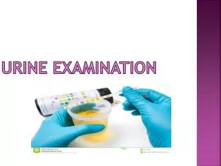
Introduction • Physical examination of urine includes • Color • Clarity • Specific gravity • Results provide • Preliminary information • Correlation with other chemical and microscopic results
Physical Characteristics • Provides preliminary information concerning disorders such as • Glomerular bleeding • Liver disease • Inborn errors of metabolism • Urinary tract infection • Renal tubular function
Color • Ranges from colorless to black • Normal variations caused by • Normal metabolic functions • Physical activity • Ingested materials • Pathologic conditions • Abnormal variations caused by • Bleeding • Liver disease • Infection
Normal Urine Color • Common terminology • Pale yellow, yellow, dark yellow • Should be consistent within institution • Urochrome is pigment causing yellow color • Normally excreted at a constant rate • Increased in thyroid disorders and fasting • Increases when specimen sits at room temperature • Provides estimate of body hydration • Pale yellow to dark yellow can be normal
Normal Urine Color (cont’d) • Additional pigments uroerythrin, urobilin • Color changes in older specimens • Uroerythrin • Pink pigment • Attaches to amorphous urates formed in refrigerated specimens • Urobilin • Oxidation of normal constituent, urobilinogen • Orange-brown color in older specimens
Abnormal Urine Color • Many colors and causes • Often reason patient comes to the physician • Common abnormal colors • Dark yellow/amber/orange • Red/pink/brown • Brown/black • Blue/green
Dark Yellow/Amber/Orange • Dark yellow and amber • Normal = concentrated urine • Abnormal = bilirubin • Bilirubin indicates possible hepatitis virus present • Standard precautions • Foam • Bilirubin produces yellow foam when shaken • Normal urine produces small amount of white foam caused by protein
Dark Yellow/Amber/Orange (cont’d) • Photooxidation of large amounts of urobilinogen produces yellow-orange urine • No yellow foam when shaken • Photooxidation of bilirubin to biliverdin produces yellow-green urine
Dark Yellow/Amber/Orange (cont’d) • Phenazopyridine (pyridium) or Azo-Gantrisin for urinary tract infection produces thick orange pigment and yellow foam (no bilirubin) • Thick pigment is noticeable, obscures natural color, and interferes with reagent strips
Red/Pink/Brown • Blood is a common cause of red urine • Color can range from pink to brown • Pink = small amount of blood • Brown = oxidation of hemoglobin to methemoglobin • Methemoglobin • RBCs remaining in acid urine • Fresh brown specimen can indicate glomerular bleeding • Cloudy red urine = RBCs • Clear red urine = hemoglobin/myoglobin • Hemoglobin • In vivo lysis of RBCs • Patient’s plasma will also be red • Consider in vitro lysis/specimen handling
Red/Pink/Brown (cont’d) • Myoglobin • Breakdown of skeletal muscle • Fresh urine is often more reddish/brown • Patient’s plasma is clear • Port wine–colored urine • Oxidation of porphobilinogen to porphyrias • Nonpathogenic red urine • Menstrual contamination • Pigmented foods • Medications (rifampin, pheno-compounds) • Fresh beets • Genetically susceptible people in alkaline urine • Black raspberries in acid urine
Brown/Black • Additional testing for specimens that • Turn black after standing at room temperature • Test negative for blood • Melanin • Excess in malignant melanoma • Oxidation of melanogen to melanin • Homogentisic acid • Black color in alkaline urine • Alkaptonuria • Medications, levodopa, phenol derivatives, flagyl
Blue/Green • Urinary and intestinal bacterial infections are the pathogenic cause • Urinary: pseudomonas infection • Intestinal: infection causing increased urinary indican oxidizing to indigo blue • Catheter bags: purple color from Klebsiella, Providencia, and indican • IV phenol medications cause green • Clorets (green) medications: Robaxin, methylene blue, Elavil (blue)
Color and Clarity Procedure • Use a well-mixed specimen • View through a clear container • View against a white background • Maintain adequate room lighting • Evaluate a consistent volume of specimen • Determine color and clarity
Clarity • Refers to the transparency or turbidity of a specimen • Normal reporting • Clear, hazy, cloudy, turbid, milky • Visual examination • Gently swirl specimen in a clear container in front of a good light source • Automated turbidity readings are available • Fresh clean-catch urine is normally clear
Urine Clarity • Clear: No visible particulates, transparent • Hazy: Few particulates, print easily seen through urine • Cloudy: Many particulates, print blurred through urine • Turbid: Print cannot be seen through urine • Milky: May precipitate or be clotted
Nonpathogenic Turbidity • Hazy female specimens with squamous epithelial cells and mucus • Bacterial growth in nonpreserved specimens • Refrigerated specimens with precipitated amorphous phosphates (white) and urates (pink) • Contamination: fecal, talc, semen, vaginal creams, IV contrast media
Pathologic Turbidity • Most common: RBCs, WBCs, bacteria • Also: nonsquamous epithelial cells, yeast, abnormal crystals, lymph fluid, lipids • The extent of turbidity should correspond to the amount of material observed in the microscopic examination • Clarity is one of the criteria considered in determining the necessity of performing a microscopic examination
Specific Gravity (SG) • Evaluation of urine concentration • Determines if specimen is concentrated enough to provide reliable screening results • Definition: the density of a solution compared with the density of an equal volume of distilled water at the same temperature
Specific Gravity (SG) (cont’d) • Isosthenuric: SG of 1.010 (the SG of the plasma ultrafiltrate) • Hyposthenuric: SG lower than 1.010 • Hypersthenuric: SG higher than 1.010 • Normal random specimen range • 1.003 to 1.035; most common 1.015 to 1.025 • Below 1.003 may not be urine • Consistent low readings: further testing
Refractometer • Measures velocity of light in air versus velocity of light in a solution • Concentration changes the velocity and angle at which the light passes through the solution • Prism in the refractometer determines the angle that light is passing through the urine and converts angle to calibrated viewing scale
Refractometer(cont’d) • Advantages • Temperature compensation not needed • Light passes through temperature-compensating liquid • Compensated between 15°C and 38°C • Small specimen size: one or two drops
Glucose and Protein Corrections • Subtract 0.003 for each gram of protein present • Subtract 0.004 for each gram of glucose present • Protein or glucose concentration can be determined from the chemical reagent strip tests
Correction Example • A specimen containing 1 g/dL protein and 1 g/dL glucose has a specific gravity reading of 1.030 • Calculate the corrected reading 1.030 – 0.003 (protein) = 1.027 – 0.004 (glucose) = 1.023 corrected specific gravity
Methodology • Drop of urine placed on prism • Focus on light source, and read scale • Wipe off prism between specimens • Calibration • Distilled water should read 1.000; adjust set screw if necessary • 5% NaCl should read 1.022 ± 0.001 • 9% sucrose should read 1.034 ± 0.001
Clinical Correlations • Abnormally high results = >1.040 • Radiographic contrast media (IVP) • Dextran, other IV plasma expanders • Check patient’s clinical course/history • Reagent strip readings and osmometry not affected by high-molecular-weight substances • Should be used as an alternative if possible
Osmolality • A more representative measure of renal concentrating ability can be obtained • Specific gravity depends on the number of particles present in a solution and the density of these particles • Osmolality is affected only by the number of particles present • Substances of interest are small molecules • Sodium • Chloride • Urea
Osmole • 1 g molecular weight of a substance divided by the number of particles into which it dissociates (= to MW of substance) • Glu = 180 g/osm (C + H + O) • NaCl = 58.5 g/osm (Na + Cl) • The unit of measure used in the clinical laboratory is the milliosmole (mOsm)
Osmolarity • Osmolality of a solution can be determined by measuring a property that is mathematically related to the number of particles in the solution • Colligative property • Changes in colligative properties • Lower freezing point • Higher boiling point • Increased osmotic pressure • Lower vapor pressure
Measuring Osmolality • Measuring osmolality in the urinalysis laboratory requires an osmometer • Additional step in the routine urinalysis procedure • Automated osmometer utilizes freezing point depression to measure osmolality
Reagent Strip SG • The reagent strip reaction is based on the change in pKa (dissociation constant) of a polyelectrolyte in an alkaline medium • Releasing H ions in direct proportion to the number of ions in the solution • The more hydrogen ions released, the lower is the pH • Indicator bromothymol-LS blue on the reagent pad measures the change in pH • Indicator changes from blue (1.000 [alkaline]), through shades of green, to yellow (1.030 [acid]) • Not affected by nonionizing substances
Odor • Not routinely reported • Fresh urine: faintly aromatic • Older urine: ammonia • Metabolic disorders: maple syrup urine disease, ketosis (fruity), infection (ammonia/unpleasant) • Food: garlic, onions, asparagus (genetic: only certain people can smell asparagus, but all produce odor)