Microscopic Examination of Urine
Microscopic Examination of Urine. Download http://www.vetlab.com/kova.htm Definition of urine sediment: all solid materials suspended in the urine - a semiquantative evaluation of the urine sediment Significance of formed elements in the urine

Microscopic Examination of Urine
E N D
Presentation Transcript
Microscopic Examination of Urine • Download http://www.vetlab.com/kova.htm • Definition of urine sediment: all solid materials suspended in the urine • - a semiquantative evaluation of the urine sediment • Significance of formed elements in the urine • Well performed microscopic exam can provide information nearly equivalent to a biopsy. • Most time consuming part of UA & until recently the least standardized. • Ongoing controversy as to when / if to perform the microscopic exam.
Microscopic Examination of Urine • Not on lecture guide. Review info in Table 6-1 • Correlation of findings from physical & chemical analysis with expectations in microscopic.
Microscopic Examination of Urine • Specimen requirements • Collection of specimen • Prefer the concentrated first morning specimen, collected = mid-stream, clean catch . • first morning most concentrated and will be able to demonstrate the most abnormalities. Mid stream, clean catch technique will eliminate fecal & vaginal contamination • Container must be clean and free of lint / debris • usually disposable plastic, must be sure no soap residue • Fresh – tested within 2 hours of voiding, or refrigeration needed.
Microscopic Examination of Urine • Obj.35. List the correct steps in the collection and preparation of a urine sample for microscopic exam. • Preparation of specimen need to standardize as much as possible • Sources of Variation (not on lecture guide) • Collection method • Centrifugation time and speed • Re-suspension of sediment • Type of microscope slide • Viscosity of specimen • Reporting of the results
Microscopic Examination of Urine • Preparation of specimen (show video) • Mix specimen well • Pour 12 ml into urine centrifuge tube • Centrifuge five minutes, 1200-2000 RPM (speed varies depending on the centrifuge’s characteristics) • Speed and time should be consistent. The “relative centrifugal force” is important.
Microscopic Examination of Urine • Pour off supernatant - except last .5-1 mL. have pipettes that assist • Re-suspend sediment - mix well, tap, or use pipette provided • Evaluate sediment in a chamber standardized for given volume and depth of field. - “In-house methods = Mount a small drop on a clean slide, cover-slip - or use commercial materials such as Count 10 • Use standardized reporting format consistent with other techs in the institution
Microscopic Examination of Urine • Commercial systems • UriSystem – slide to follow • KOVA System – video or several slides to follow • Count -6 or Count 10 • all have their ‘own brand’ of tubes, pipettes, stain, slides, etc. • Authors also mentions several other ‘all in one-type of systems’
Microscopic Examination of Urine • UniSystem Standardization of Urine Sediment
Microscopic Examination of Urine • Sedi-Stain (Sternheimer and Malbin) crystal violet, safranin-O • Sedi-Stain & KOVA stain are commercial preparations with addition of stabilizers to prevent precipitation. • Supra-vital stain used to increase visibility of structures. Assists greatly in differentiating renal tubular epithelial cells (which will take on an eosinophilic - oranges cytoplasm & dk purple nuclei) from transitional epithelial (which are more over-all blue)
Microscopic Examination of Urine • Not on lecture guide – Table 6-3 • Sediment stain characteristics • Toluidine blue – nuclear structure • Assists in differentiating WBC from renal epith. • 2% acetic acid - removes interfering RBCs and enhances nuclei of WBC • Lipid stains - Oil Red O, Sudan III - stains triglycerides and neutral fats orange-red to ID lipid containing cells.
Microscopic Examination of Urine • Gram stain - to assist in ID of gram reaction of bacteria. • Hansel stain - methylene blue and eosin Y stains eosinophilic granules - ID eosinophils • Prussian blue reaction - makes iron granules blue in color (hemosiderin granules appear yellow until stained)
Microscopic Examination of Urine • Table 6-5 – page 73 provides information on types of microscopic techniques that have application in UA Brighfield microscope – very subdued light: lowered condenser, closed iris diaphragm, use filters • Continuously focus up and down with fine adjustment as you learned in hematology. • Polarized light - may use to ID crystals, lipids
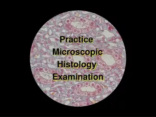
Microscopic Examination of Urine • Types of Sediment • As one author puts it: • Cells • Casts • Crystals • Critters
Microscopic Examination of Urine • Types of Sediment • Organized – biological part • RBC • WBC • Casts • Epithelial cells • Bacteria, parasites, yeast and fungi • Unorganized • Crystals • Amorphous crystalline matter.
Microscopic Examination of Urine • Examination • - should correlate with physical and chemical dipstick, may need to recheck • Scanning - – 10-15 fields using low power (10X). Look for casts, mucous, and squamous epithelial cells in general getting an overall feel • Report squamous epithelial cells, crystals, mucous, etc. using semi-quantitative terms such as rare, few, moderate, or many (or trace, 1+,2+,3+, & 4+) according to lab protocol.
Microscopic Examination of Urine • Enumeration - quantitate. Method may vary from lab to lab • Average number of RBC/hpf • Average number of WBC/hpf • Average number of any renal tubular or transitional epithelial cells /hpf.
Microscopic Examination of Urine Average number (and type) of casts/__average # of casts /hpf______ • authors have varied back and forth as whether low or high power should be reported... use low power to locate and enumerate the various types , but may need to switch to high power to identify the type... • Strasinger says report / lpf (use hpf to ID) • Unorganized sediment – few, moderate, many, packed; kinds seen • Note presence of bacteria, yeasts, crystals, epithelial cells (covered), etc. • quantitate these also
Microscopic Examination of Urine • .Changes in urine sediment when allowed to stand • important to keep in mind the changes in microscopic structures that can occur (don’t forget the other chemical changes ie bilirubin, pH, ketones) • RBC distorted – crenation, swelling, disintegration • WBC disintegrates in alkaline urine • Cast disintegrate in alkaline urine • Bacterial growth – increased alkalinity • Increased precipitation of crystals, especially amorphous
Microscopic Examination of Urine • Microscopic sediment • Red Blood Cells • White Blood Cells • Epithelial Cells • Casts • Crystals • Miscellaneous structures • Students go to end of area’s lecture guide. Continue to next slide.
Microscopic Examination of Urine • Addis Count – Strasinger page 68 • Early way of accurately enumerating urine sediment. • Actual enumeration of casts, RBC, WBC, using a hemacytometer • developed as a way to standardize urine microscopics to monitor known cases of renal disease. • Rarely done today as most urine microscopic systems produce standardized results if manufacture directions are followed.