Download
1 / 45
450 likes | 466 Vues
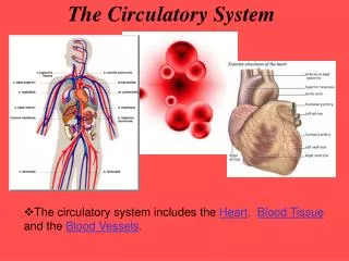
Delve into the intricate structure and functions of blood vessels, from arteries to capillaries to veins, in the body's circulatory system. Understand the roles of tunics in vessel walls and the vital process of gas exchange in capillaries.

E N D
Blood Vessels • Delivery system of dynamic structures that begins and ends at the heart • Arteries: carry blood away from the heart; oxygenated except for pulmonary circulation • Capillaries: contact tissue cells and directly serve cellular needs • Veins: carry blood toward the heart except for pulmonary circulation
Arterial system Venous system Large veins (capacitance vessels) Heart Elastic arteries (conducting vessels) Large lymphatic vessels Lymph node Muscular arteries (distributing vessels) Lymphatic system Small veins (capacitance vessels) Arteriovenous anastomosis Lymphatic capillary Sinusoid Arterioles (resistance vessels) Postcapillary venule Terminal arteriole Metarteriole Thoroughfare channel Precapillary sphincter Capillaries (exchange vessels) Figure 19.2
Structure of Blood Vessel Walls • Arteries and veins • Composed of layers called “tunics” • (3) Tunica intima, tunica media, and tunica externa • Lumen • Central blood-containing space • Capillaries • Endothelium with sparse basal lamina
Tunica intima Valve • Endothelium • Subendothelial layer Internal elastic lamina Tunica media (smooth muscle and elastic fibers) External elastic lamina Tunica externa (collagen fibers) Lumen Vein Lumen Artery Capillary network Basement membrane Endothelial cells Capillary (b) Figure 19.1b
Tunic = covering • Tunica intima • Endothelium lines the lumen of all vessels • In vessels larger than 1 mm, a subendothelial connective tissue basement membrane is present Tunics
Tunics • Tunica media • Smooth muscle and sheets of elastin • Sympathetic vasomotor nerve fibers control vasoconstriction and vasodilation of vessels
Tunics • Tunica externa (tunica adventitia) • Collagen fibers protect and reinforce • Larger vessels contain vasa vasorum to nourish the external layer
Arterial System Divided into 3 groups based on size and function • Elastic (Conducting) Arteries • Muscular (Distributing) Arteries • Arterioles
Elastic (Conducting) Arteries • Large thick-walled arteries with elastin in all three tunics • Aorta and its major branches • Large lumen offers low-resistance • Act as pressure reservoirs—expand and recoil as blood is ejected from the heart
Muscular (Distributing) Arteries and Arterioles • Distal to elastic arteries; deliver blood to body organs • Have thick tunica media with more smooth muscle • Active in vasoconstriction
Arterioles • Smallest arteries • Lead to capillary beds • Control flow into capillary beds via vasodilation and vasoconstriction
Capillaries • Microscopic blood vessels • Only blood vessels to permit gas exchange • Very thin walls only one cell thick • Small diameter allows only a single RBC to pass at a time
Capillaries • In all tissues except for cartilage, epithelia, cornea and lens of eye • Functions: exchange of gases, nutrients, wastes, hormones, etc.
Arterial system Venous system Large veins (capacitance vessels) Heart Elastic arteries (conducting vessels) Large lymphatic vessels Lymph node Muscular arteries (distributing vessels) Lymphatic system Small veins (capacitance vessels) Arteriovenous anastomosis Lymphatic capillary Sinusoid Arterioles (resistance vessels) Postcapillary venule Terminal arteriole Metarteriole Thoroughfare channel Precapillary sphincter Capillaries (exchange vessels) Figure 19.2
Capillary Beds • Interwoven networks of capillaries form the microcirculation between arterioles and venules
Capillary Beds Consist of two types of vessels • Vascular shunt (metarteriole—thoroughfare channel): • Directly connects the terminal arteriole and a postcapillary venule • True capillaries • 10 to 100 per capillary bed depending on organ or tissue being served • Branch off the metarteriole or terminal arteriole
Blood Flow Through Capillary Beds • Precapillary sphincters regulate blood flow into true capillaries • Regulated by local chemical conditions and vasomotor nerves • Sphincters are open or closed depending on organ or tissue needs at the time ex. After a meal, sphincters are open to increase blood flow to aid in digestion. Otherwise, they are closed due to decreased demand
Vascular shunt Precapillary sphincters Thoroughfare channel Metarteriole True capillaries Terminal arteriole Postcapillary venule (a) Sphincters open—blood flows through true capillaries. Terminal arteriole Postcapillary venule (b) Sphincters closed—blood flows through metarteriole thoroughfare channel and bypasses true capillaries. Figure 19.4
Venules • Formed when capillary beds unite • Very porous; allow fluids and WBCs into tissues • Postcapillary venules consist of endothelium and a few pericytes • Larger venules have one or two layers of smooth muscle cells
Veins • Formed when venules converge • Still have 3 tunics but have thinner walls & larger lumens compared with corresponding arteries • Blood pressure is lower than in arteries • Thin tunica media and a thick tunica externa consisting of collagen fibers and elastic networks • Called capacitance vessels (blood reservoirs); contain up to 65% of the blood supply
Vein Artery (a) Figure 19.1a
Pulmonary blood vessels 12% Systemic arteries and arterioles 15% Heart 8% Capillaries 5% Systemic veins and venules 60% Figure 19.5
Veins • Adaptations that ensure return of blood to the heart • Large-diameter lumens offer little resistance • Valves prevent backflow of blood • Most abundant in veins of the limbs
Homeostatic Imbalance Vericose Veins • Caused by leaky valves that dilate the vein • Pooling blood over time weakens the valves and stretches the venous walls • Usually affects the lower limbs
Homeostatic Imbalance • Vericose Veins • Contributing factors include: • Heredity • Prolonged standing • Obesity • Pregnancy • Increased pressure from bearing down during child birth or bowel movements can cause vericosities known as hemorrhoids
Physiology of Circulation: Definition of Terms • Blood flow • Volume of blood flowing through a vessel, an organ, or the entire circulation in a given period • Measured as ml/min • Equivalent to cardiac output (CO) for entire vascular system • Relatively constant when at rest • Varies widely through individual organs, based on needs
Physiology of Circulation: Definition of Terms • Blood pressure (BP) • Force per unit area exerted on the wall of a blood vessel by the blood • Expressed in mm Hg • Measured as systemic arterial BP in large arteries near the heart • The pressure gradient provides the driving force that keeps blood moving from higher to lower pressure areas
Physiology of Circulation: Definition of Terms • Resistance (peripheral resistance) • Opposition to flow • Measure of the amount of friction blood encounters • Generally encountered in the peripheral systemic circulation • Three important sources of resistance • Blood viscosity • Total blood vessel length • Blood vessel diameter
Resistance • Factors that remain relatively constant: • Blood viscosity • The “stickiness” of the blood due to formed elements and plasma proteins • Blood vessel length • The longer the vessel, the greater the resistance encountered
Resistance • Frequent changes in diameter alter peripheral resistance • The smaller the tube, the greater the friction because more fluid contacts the tube wall • E.g., if the radius is doubled, the resistance is reduced
Resistance • Small-diameter arterioles are the major determinants of peripheral resistance • Abrupt changes in diameter or fatty plaques from atherosclerosis dramatically increase resistance • Disrupt flow and cause turbulence • If resistance increases, blood flow decreases
Systemic Blood Pressure • The pumping action of the heart generates blood flow • Pressure results when flow is opposed by resistance • Systemic pressure • Is highest in the aorta • Declines throughout the pathway • Is 0 mm Hg in the right atrium • The steepest drop occurs in arterioles
Systolic pressure Mean pressure Diastolic pressure Figure 19.6
Arterial Blood Pressure • Reflects two factors of the arteries close to the heart • Elasticity (compliance or distensibility) • Volume of blood forced into them at any time • Blood pressure near the heart is pulsatile
Arterial Blood Pressure • Systolic pressure: pressure exerted during ventricular contraction • Diastolic pressure: lowest level of arterial pressure • Pulse pressure = difference between systolic and diastolic pressure
Capillary Blood Pressure • Ranges from 15 to 35 mm Hg • Low capillary pressure is desirable • High BP would rupture fragile, thin-walled capillaries • Most are very permeable, so low pressure forces filtrate into interstitial spaces
Venous Blood Pressure • Changes little during the cardiac cycle • Small pressure gradient, about 15 mm Hg • Low pressure due to cumulative effects of peripheral resistance
Factors Aiding Venous Return • Respiratory “pump”: pressure changes created during breathing move blood toward the heart by squeezing abdominal veins as thoracic veins expand • Muscular “pump”: contraction of skeletal muscles “milk” blood toward the heart and valves prevent backflow • Vasoconstriction of veins under sympathetic control
Valve (open) Contracted skeletal muscle Valve (closed) Vein Direction of blood flow Figure 19.7
Maintaining Blood Pressure • Requires • Cooperation of the heart, blood vessels, and kidneys • Supervision by the brain
Maintaining Blood Pressure • The main factors influencing blood pressure: • Cardiac output (CO) • Peripheral resistance (PR) • Blood volume
Maintaining Blood Pressure • F = P/PR and CO = P/PR • Blood pressure = CO x PR (and CO depends on blood volume) • Blood pressure varies directly with CO, PR, and blood volume • Changes in one variable are quickly compensated for by changes in the other variables