Acetylcholine (ACh)
Acetylcholine (ACh). Acetylcholine Serotonin Dopamine Noradrenaline. E13, SN, VTA neocortex innervation E15. Herlenius, Lagercrantz; 2001,2004. Ionotróp receptorok: ligandum-vezérelt ioncsatornák. N. N. C. GABA A nAChR 5HT-3 GlyR. P2X. GluR. C. C. N.
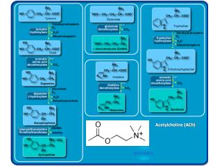

Acetylcholine (ACh)
E N D
Presentation Transcript
Acetylcholine Serotonin Dopamine Noradrenaline E13, SN, VTA neocortex innervation E15 Herlenius, Lagercrantz; 2001,2004
Ionotróp receptorok: ligandum-vezérelt ioncsatornák N N C GABAAnAChR5HT-3GlyR P2X GluR C C N
GABA-vezérelt ioncsatornák: GABAA receptorok g-amino-vajsav, 4-aminobutirát g-amino-butyric acid GABA: „ősi” parakrin szabályozó hatóanyag HOOC-CH2-CH2-CH(NH2)-COOH → CO2 + HOOC-CH2-CH2-CH2NH2 Bioszintézis: GAD65, GAD67 Glutaminsav-dekarboxiláz Glutamic acid decarboxylase + piridoxálfoszfát Felvétel, akkumuláció: GABA-transzporterek Vezikuláris GABA-transzporter glutaminsav GABA + CO2 lebontás: GABA-transzamináz (citrát-körben lebontás) Ionotróp (GABAA) és metabotróp (GABAB) receptorok
Ionotróp GABA receptorok (GABAA) Alegységek (emberben): 6α (GABRA1, GABRA2, GABRA3, GABRA4, GABRA5, GABRA6) 3 β (GABRB1, GABRB2, GABRB3) 3 γ (GABRG1, GABRG2, GABRG3) δ (GABRD), ε (GABRE), π (GABRP θ (GABRQ) 3 ρ (GABRR1, GABRR2, GABRR3 (GABAC receptorok; retina) ([http://www.tocris.com/pdfs/gabarev.pdf]
GAD67 P1 rat CNS fejlődésének minden szakaszában Popp et al., 2009
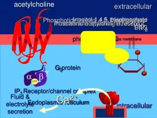
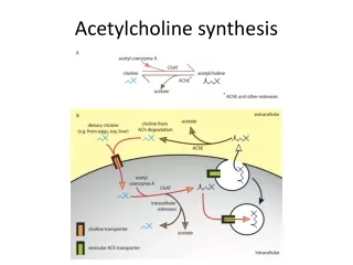
Gyors deszenz. Hyperpol mp., depol Acetilkolin-vezérelt ionotróp receptorok (nAchR)
AMPA-receptorok α-amino-3-hydroxy-5-methyl-4-isoxazolepropionic acid Q: glutamine R : arginine GluR2 Ionotróp glutamát receptorok
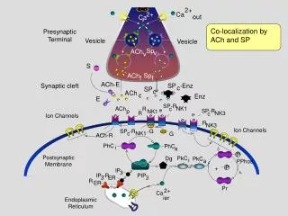
Expression and distribution of iGlu receptors and GABAB receptors in CP cells and tangentially migrating interneurons. CP cells may release glutamate that could activate (blackpositive symbol) iGlu receptors expressed in the tangentially migrating interneurons. This activation may lead to GABA release from these cells andto the activation of its GABA receptors and those expressed in nearby cells. The blockade of GABAB receptor leads to an accumulation of interneuronsat the proliferative zones of the cortex suggesting that the activation of this receptor is important for the transition of the interneurons from the LIZ to the CP of the cortex. MZ, marginal zone. Neurotransmitter receptors are involved in the Prolif and migration of cortical neurons. AMPA responses are first observed in terminally dividing neuronal progenitors while postmitotic neurons (green cells) express both AMPA/kainate and NMDA receptors. Activation of GABA and Glu receptors by GABA and Glu, respectively, has been shown to shorten the cell cycle of VZ progenitors (black circled positive symbol), while the Prolif in the SVZis markedly decreased in response to GABA (black circled negative symbol). Neurotransmitter receptor activation has been reported to influenceneuronal migration of cortical neurons at early stages of development. Thus, GABAC and GABAB-like receptor activation (red circled positive symbol)stimulates migration of neurons from the VZ and IZ, respectively, whereas GABAA receptor activation arrests migration as neurons approach theirtarget destinations in the CP. The U-shape discontinuous line with arrows represents the direction followed by the cells types during Prolif andmigration. The gradient in the background color represents the different cortical layers. Glu, glutamate; MZ, marginal zone; Prolif, proliferation; RG, radial glia. R. Luján et al. / Neuroscience 130 (2005) 567–580