Download
1 / 25
250 likes | 430 Vues
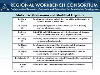
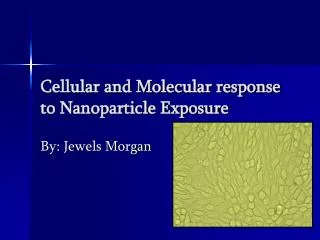
Cellular and Molecular response to Nanoparticle Exposure. By: Jewels Morgan. What is a nanoparticle?. Small particles with a dimension of less than 100nm. Nanoparticles Uses. Used in many different applications Biomedical Optical Electronics Hygiene Products Safety Products

E N D
Cellular and Molecular response to Nanoparticle Exposure By: Jewels Morgan
What is a nanoparticle? • Small particles with a dimension of less than 100nm
Nanoparticles Uses • Used in many different applications • Biomedical • Optical • Electronics • Hygiene Products • Safety Products • Food Industry
What are possible dangers of Nanoparticle Exposure? • Cross biological membrane • Small surface area/volume ratio • Toxicity to human tissues and cell cultures • Possible DNA interactions, damage, mutations • Uptake by mitochondria and possible damage • Cell death
Previous Research • Cultured two different cell lines • CHO (Chinese Hamster Ovary) • COS-7 (African Green Monkey Kidney Cells) • Morphology of both cell lines • Cytotoxicity of both cell lines • Bead location • Preliminary Apoptosis
Particles Used • Latex Beads, Blue dyed • Ultra fine nanoparticles (55nm) • Fine particles (800nm)
Fall 2007 Research Results • Cytotoxicity Results • CHO 24 hrs • COS-7 48 hrs • Both bead sizes showed cytotoxic affects to cells • Morphology • Cultured cells morphology matched ATCC database • Nanoparticle Location • Determined localization of the nanoparticles to be in the cytoplasmic region
Pictures of Research Results Bead Location CHO 24 hr exposure Morphology
Current Research Spring 2008 • Apoptosis • CHO cells • CASP1 (Caspase) • Chromosomal Spread-Aberrations • Halted in metaphase using colchicine • Gene Expression of Pro-Inflammatory Cytokines-PCR RNA • IL-6 • IL-18 • Protein Concentration
Apoptosis • Programmed Cell death • CHO Cells • Treated for 24 hrs with 1μL of beads • Isolated DNA • Verified purity and concentration • Spectrophotometer • λ260nm and λ280nm
DNA Purity All values determined using a 1:100 dilution Ratio value 1.60 to 1.80=pure DNA
Showed no fragmentation of DNA No significant evidence of Apoptosis occurring DNA Analysis following nanoparticle treatment Ladder 55nm CHO DNA
Pro-Inflammatory Cytokines • Isolated RNA • TRI Reagent • Lyse cells to release RNA • RNA Isolated from Controls, 55nm, & 800nm exposure cells • PCR samples with designed primers for: • IL-6 • IL-18 • Caspase • Beta Actin
Pro-Inflammatory Cytokines and Apoptotic gene • IL-6 • Released following oxidative stress to cell • IL-18 • Released following oxidative stress to cell • CASPASE • Elevated during Apoptosis
Primer Design • NCBI Database • Chinese Hamster (Cricetulus griseus) IL-6 • Mouse (Mus musculus) Caspase -1 • Unknown Sequence in CHO • Mouse (Mus musculus) IL-18 • Unknown Sequence in CHO • Chinese Hamster (Cricetinae gen. sp) Beta Actin • Endogenous gene expression
Primer Design cont. • Primers need to have: • 50% or greater gc content • Annealing temperature ~60.0ºC • Product size ~350-450bp • Primers designed using Primer 3 software after importing sequence from NCBI database • Primer size 20bp
RNA Purity and Concentration Results All values determined using a 1:100 dilution Ration value of 1.8-2.0=Pure RNA
Gene Expression Ladder Ladder Con 55nm 800nm Con 55nm 800nm 1000bp Ladder 500bp 1000bp 500bp CHO IL-6 gene CHO CAS gene
Pro-Inflammatory Cytokine Response cont. Ladder Control 55nm 800nm Ladder Control 55nm 800nm IL-18 Beta Actin
Chromosomal Spread-Aberrations Control CHO Chromosomes 55nm Chromosome Spread 800nm Chromosome Spread
* * Significantly different from control student’s t-test
Summary • DNA Apoptosis • No detection of Apoptotic bands • Increase in Caspase expression • Cytokine Expression • IL-6 increased following treatment with 55nm beads compared to control • IL-18-No detection of gene expression • Slight protein concentration in treated samples • Chromosome number and appearance altered following treatment
Special Thanks to…. • Dr. Jacqueline Jordan-Research Mentor • Truyen Derek Pham-Supplies Donation • Emory University • Sheryl Sands-Lab Technician • Dr. Michelle Furlong • CSU Natural Sciences Faculty & Staff