Download
1 / 25
250 likes | 373 Vues
Phases of Healing. Sally white, MOTS The university of M ississippi medical center. Phases of Healing. There are 3 main phases of healing for soft tissue Inflammatory Phase Typically begins within the first 6-8 hours Time frame: 2-5 days

E N D
Phases of Healing Sally white, MOTS The university of Mississippi medical center
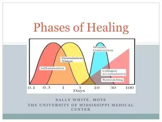
Phases of Healing • There are 3 main phases of healing for soft tissue • Inflammatory Phase • Typically begins within the first 6-8 hours • Time frame: 2-5 days • Macrophages become the dominate cell type to debride damaged tissue • Proliferation Phase • Fibroblast cells mark the beginning of this phase • Lay down at collagen matrix • Site appears red due to need for oxygen • Collagen deposition occurs • Remodeling/Maturation Phase • Collagen production and degradation equalize • Strength approaches about 50% of tissue’s normal strength about 3 months post injury All phases over-lap
Patient Factors Affecting Healing • Diabetes Mellitus • Diabetic scars have less collagen • Collagen that is laid down is more brittle than normal • May not have good blood supply; especially to distal extremities • Malnutrition • Slow healing due to breakdown of protein source for energy • May have vitamin deficiency • Age • Increase in age is an increase in healing time • Smoking • Infection to Site/ Re-injury • Local Skin Tension • Ischemia/Hypoxia
Tendons • Are cord-like structures that attach muscles to bones • Can be classified as gliding or vascular • Gliding: Enclosed in the tendon sheath- the synovial sheath • Common example: Flexor tendons in the Hand • Vascular: Are surrounded by connective tissue • Achilles Tendon
Tendons Tendons are stronger per unit area than muscle, and their tensile strength equals that of bone, although it is flexible and slightly extensible. Collagen constitutes 75% of the dry tendon weight and functions chiefly to withstand and transmit large forces between muscle and bone.
Common Tendon Injuries • Common Injuries: • Tendonitis- Inflammation • Common areas: RTC (supraspinatus), lateral epicondylitis, patellar tendon • Tendinosis- Degenerative • Chronic, accompanied by pain and associated with thickening of the tendon itself • Paratenonitis- Inflammatory disorder of tissue surrounding the tendon (sheath) • Common cause: Repetitive friction • Also goes by: Peritendonitis, Tenosynovitis
Tendon Phases of Healing • Initial Inflammatory phase • Begins within 24 hours of injury • Neutrophils enter injury site • Tenocytes migrate to wound gradually for type-III collagen synthesis initiation • Therapy during this phase: • Controlled stretch to increase collagen synthesis and to improve fiber alignment. Gentle passive range of motion can result in higher tensile strength. • Repetitive motion can increase DNA content and increase protein synthesis. • Proliferative Phase • Last up to 6 weeks • Tendon fibroblast synthesizing collagen and other matrix components • Synthesis of type III collagen peaks- water content remains high
Tendon Phases of Healing Continued • Remodeling Phase • Week 6- Up to 1 year post injury • Broken down into 2 stages • Consolidation- Week 6-10 • Type-I collagen • Collagen fibers align in the direction of stress response • Maturation- Week 10+ • Gradual change of fibrous tissue to scar-like tendon tissue over the course of a year
Ligaments Ligaments are the most frequently injured tissues within a joint. When a ligament is overloaded, or exposed to tensions greater than the structure can sustain, the tissue fails, resulting in partial or complete ligament discontinuities, more commonly known as disruptions or tears.
Ligaments Ligament injures create disruptions in the balance between joint mobility and stability, causing abnormal force transmission, which results in damage to other structures in and around the joint. The long-term consequence of non-healed ligament injury is osteoarthritis, the most common joint disorder. Ligaments heal through a distinct sequence of cellular events that take place in three consecutive stages.
Ligament Phases of Healing • Acute inflammatory phase • Begins within minutes of injury and continues over the next 48 to 72 hours. • Blood collects at the site of injury and platelet cells interact with certain matrix components, changing their shape and initiating clot formation. • When stimulated by growth factors, neutrophils, monocytes, and other immune cells migrate to the injured tissue where they ingest and remove debris and damaged cells: thereby initiating matrix turnover.
Ligament Phases of Healing • Proliferative phase • Begins when immune cells release various growth factors and cytokines: which initiates fibroblast proliferation signals for rebuilding of the ligament tissue matrix. • The tissue formed initially appears as disorganized scar tissue, consisting of more blood vessels, fat cells, fibroblasts, and inflammatory cells than normal ligament tissue contains. • Over the next several weeks, fibroblast cells deposit various types of collagen, proteoglycans, glycoproteins, and other proteins into the matrix. • The collagen becomes aligned with the long axis of the ligament during this time; however, the newly-formed collagen fibrils are abnormal and smaller in diameter than normal ligament tissue.
Ligament Phases of Healing • Remodeling phase • Collagen maturation begins, often lasting for months to as long as years after the initial injury. • The tissue matrix starts to resemble normal ligament tissue; however, critical differences in matrix structure and function persist. • Evidence suggests that the injured ligament structure is replaced with tissue that is grossly, histologically, biochemically, and biomechanically similar to scar tissue
Bone heals with bone- which is unlike any other tissue which heals with scar tissue Type of healing (primary or secondary) depends on method of treatment of the fracture Bone Healing
Primary Bone Healing/Direct or Primary Cortical The cortex of the bone attempts to directly re-establish continuity Rigid stabilization/Fixation Some regions of the cortex are in direct contact with other cortical region at fracture site There is no callus formation
Primary Healing Contact Healing Gap Healing Healing takes place across contact area (<0.01mm) Bone reconstruction begins with direct formation of lamellar bone Initiated by formation of “cutting cones” close to fracture line When small gaps (1mm) form between bone ends and interfragmentary deformation is very low Healing involves the osteoblast cells to invade blood vessels- concentric bony rings around Haversian systems & osteoclasts to remodel
Secondary Bone Healing This is how the majority of bones heal Also called indirect bone healing Relies on the periosteum for the healing process The amount of callus formation depends on the stability of the fracture
Phases of secondary bone healing • Impaction Phase • Bone absorbs the energy; fractures along the line of least resistance • Impact damages the bone, bone marrow, periosteum, adjacent soft tissue, and vessels within the bone • Inflammatory Phase • Begins immediately after initial disruption and last 3-4 days • Hematoma forms- which is critical to healing. It allows for structural stability and is the framework for the new bone • Thrombosis occurs • Pain, swelling and heat at fracture site
Secondary Bone Healing • Repair Phase • Last about 2 weeks • Growth of new capillaries • Slight gain in mechanical strength of fracture • Soft callus formation • Callus Mineralization Stage • Begins 1 week after soft callus formation. This stage can last 4-16 weeks • Increased oxygen tension • The amount of callus is dependent on stability of fracture fragments • The more motion at the fracture site will result in larger callus formation • Initiation of controlled AROM can begin between 3 and 6 weeks post-injury if the fixation of the fracture is adequate
Bone Healing • Remodeling Phase • Can take several years to complete • Bone continues to form and becomes more compact • Dependent on adequate blood supply and stability
Wounds/Burns • Inflammatory • Last 3-5 Days • Bacteria is phagocytosed and removed from site • Begin the cleaning of the site • First Part of Inflammatory Phase: Vascular response • Main task: stop hemorrhage • Second Part: Cellular Response • Fibrin and other proteins act as glue for wound site • Therapy: Edema reduction, wound care and maintenance, early range of motion, positioning and splinting
Wounds/Burns • Proliferative • Angiogenesis, collagen deposition, formation of granulation tissue, epithelialization, and wound contraction • Fibroblast arrive to further strengthen the wound • Therapy: Edema reduction, Wound care and maintenance, early range of motion, positioning and splinting, stretching and strengthening
Wounds/Burns • Remodeling • Collagen is remodeled and realigned along the tension lines • Scar maturation takes 6-18 months • This stage is fragile and can take up to 2 years for a severe burn patient • Therapy:Range of motion, strengthening and reconditioning, scar management techniques
References Hauser, R.A., Dolan, E.E., Phillips, A.C., Moore, R.E., & Woldin, B.A. Ligament Injury and healing: a review of current clinical diagnostics and therapeutics. The Open Rehabilitation Journal, 2013, 6, 1-20. McCulooch, J.M. &Kloth, L.C. (year), Rehabilitation of the burned individual. In Wound Healing- Evidence Based Management (4 ed, pp. 357-390) Pendleton, H.M. & Schultz-Krohn, W. Pedretti’s Occupational Therapy Practice Skills for Physical Dysfunction (6th edition, 994-995, 1005-1007, 1022-1024) Sharma, P. & Maffulli, N. Tendon injury and teniopathy: healing and repair. J Bone Joint Surg. Am. 87:187-202, 2005. doi: 10.2106/JBJ5.D.01850.