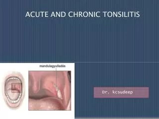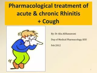
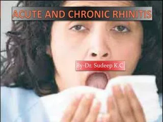
ACUTE AND CHRONIC RHINITIS
ACUTE AND CHRONIC RHINITIS. By-Dr. Sudeep K.C. 1) VIRAL RHINITIS. 1)COMMON COLD(CORYZA) Aetiology : it is caused by virus usually through airborne droplets. Adeno virus , picorna virus, rhino and coxsackie virus. Incubation period is 1-4 days and illness last for 2-3 weeks.

ACUTE AND CHRONIC RHINITIS
E N D
Presentation Transcript
ACUTE AND CHRONIC RHINITIS By-Dr. Sudeep K.C.
1) VIRAL RHINITIS 1)COMMON COLD(CORYZA) Aetiology: it is caused by virus usually through airborne droplets. • Adeno virus , picorna virus, rhino and coxsackie virus. • Incubation period is 1-4 days and illness last for 2-3 weeks.
CLINICAL FEATURES: • Burning sensation of nose followed by nasal stuffiness , rhinorrhoea and sneezing. • Low grade fever. • Nasal discharge is initially watery and profuse but may become mucopurulent due to sec. bacterial invasion. • TREATMENT: • Bed rest • Plenty of fluids. • Antihistamine and nasal decongestant. • Analgesics to relieve headache and antibiotics if secondary infection.
COMPLICATIONS: • Disease is usually self limiting and resolves spontaneously after 2to 3 weeks. • Occasionally sinusitis, bronchitis, pharyngitis may occur. INFLUENZAL RHINITIS: • Caused by influenza viruses A , B or C . RHINITIS ASSOCIATED WITH EXANTHEMAS: • Measles, rubella, chickenpox, are often associated with rhinitis which precedes exanthemas by 2-3 days.
2) BACTERIAL RHINITIS A)Non-specific infections: • It may be primary or secondary. • Primary bacterial rhinitis is seen in child usually infected by pneumococcus , streptococcus or staphylococcus. • A greyish white membrane may form in the nose , which with attempted removal, cause bleeding. B)Secondary bacterial rhinitis is result of bacterial infection supervening acute viral rhinitis.
3) IRRITATIVE RHINITIS • Caused by exposure to dust, smoke and irritating gases like ammonia, formalin etc. • May result from trauma on nasal mucosa during intranasal manipulation. CLINICAL FEATURES: • Immediate catarrhal reaction with sneezing, rhinorrhoea and nasal congestion. • Symptoms may pass off rapidly with removal of offending agent .
CHRONIC SIMPLE RHINITIS Aetiology: • Recurrent attacks of acute rhinitis in presence of predisposing factors leads to chronicity. Predisposing factors: • Persistence of nasal infection due to sinusitis , tonsillitis and adenoids. • Chronic irritation from dust, smoke etc • Nasal obstruction due to DNS, synechia leading to persistence of discharge.
PATHOLOGY: • There is hyperaemia and edema of mucous membrane with hypertrophy of seromucinous glands and increase in goblet cells. • Blood sinusoids over turbinates are distended. CLINICAL FEATURES: • Nasal obstruction • Nasal discharge • Headache • Swollen turbinates- pit on pressure ,shrink with decongestant. • Post nasal discharge.
TREATMENT: • Treatment of causative agent. • Nasal irrigation with alkaline solution . • Nasal decongestant help to relieve nasal obstruction and improves sinus ventilation. • A short course of systemic steroids helps to wean patient already addicted to excessive use of decongestant drops or sprays. • Antibiotics.
HYPERTROPHIC RHINITIS: • It is characterised by thickening of mucosa, submucosa, seromucinous glands, periosteum, and bone. Changes are more marked on the turbinates. AETIOLOGY: • Recurrent nasal infections. • Chronic sinusitis , chronic irritation of nasal mucosa due to smoking and other irritants • Prolonged use of nasal drops and vasomotor and allergic rhinitis.
SYMPTOMS: • Nasal obstruction is main symptom. • Nasal discharge is thick and sticky. • Headache and transient anosmia. EXAMINATION: • Hypertrophy of turbinates. • Turbinate mucosa is thick and does not pit on pressure. Little shrinkage with vasoconstrictor due to underlying fibrosis.
TREATMENT: • First is to discover the cause and remove it. • Nasal obstruction can releived by reduction in size of turbinates by various methods.
ATROPHIC RHINITIS(OZAENA) • It is the chronic inflammation of nose characterised by atrophy of nasal mucosa and turbinate bones . The nasal cavities are roomy and full of foul-smelling crusts. • Two types • Primary atrophic rhinitis • Secondary atrophic rhinitis.
PRIMARY ATROPHIC RHINITIS AETIOLOGY: (HERNIA) • Hereditary factors. • Endocrinal disturbance. • Racial factors . • Nutritional deficiency. • Infective. • Autoimmune process.
PATHOLOGY: • Ciliated columnar epithelium is lost and is replaced by stratified squamous type. • Atrophy of seromucinous glands, venous blood sinusoids and nerve element. • Turbinate undergoes resorption causing widening of nasal chambers.
CLINICAL FEATURES SIGN & SYMPTOMS • Common in females during puberty. • Foul smell from nose , but patient remains unware. • Marked anosmia(merciful anosmia) • Nasal obstruction inspite of wide nasal chambers due to large crust formation. • Epistaxis.
EXAMINATION shows nasal cavity to be full of greenish or greyish black dry crusts covering the turbinates & septum. • Attempt to remove my cause bleeding. If removed , nasal cavities appear roomy with atrophy of turbinates so much so that the posterior wall of nasopharynx can be easily seen . • Nasal turbinates may be reduced to mere ridges. • Nasal mucosa appears pale . • Nasal vestibule may be present shows saddle defromity • Atrophic changes may be seen in pharyngeal mucosa larynx with cough and hoarseness of voice.
Radiographic Findings • Mucoperiosteal thickening of the paranasal sinuses. • Loss of definition of the OMC secondary to resorption of the ethmoid bulla and uncinate process. • Hypoplasia of the maxillary sinuses. • Enlargement of the nasal cavities with erosion and bowing of the lateral nasal wall. • Bony resorption and mucosal atrophy of the inferior and middle turbinates.
PROGNOSIS • The disease persists for years but there is a tendency to recover spontaneously in middle age.
Current Therapies • Goals of therapy • Restore nasal hydration • Minimize crusting and debris • Therapy options • Topical therapy • Saline irrigations • Antibiotic irrigations • Systemic antibiotics • Implants to fill nasal volume • Closure of the nostrils
Local therapy • Irrigations Saline • Mixtures • Sodium bicarbonate • Shehata: Sodium Carbonate 25g, Sodium Biborate 25g, and Sodium Chloride 50g in 250ml water. • Antibiotic solution • Moore: Gentamycin solution 80mg/L • Anti-drying agents • Glycerine • Mineral Oil • Paraffin with 2% Menthol • Other • Acetylcholine • Pilocarpine
Systemic therapy • Oral antibiotics • Tetracycline • Ciprofloxacin • Aminoglycosides • Streptomycin injections • Medication avoidance • Vasoconstrictors • Topical steroids * • Other • Vitamin A (12,500 to 15,000 Units daily) • Potassium Iodide (Increases nasal secretions) • Vasodilators • Iron therapy • Estrogen • Corticosteroids * • Vaccines • Antibacterial (Pasturella, Bordetella) • Autogenous
SURGICAL: A) Young’s operation: • Both the nostrils are closed completely just within the nasal vestibule by raising flaps. They are opened after 6 months or later. In these cases, mucosa may revert to normal and crusting reduced. • Young’s procedure • Circumferential flap elevation 1 cm cephalic to the alar rim. • Sutures placed in center of elevated flap to close the nostril • Advantages • Often provided relief of symptoms • Disadvantages • Difficult to elevate circumferential flap • Breakdown of central suture area common • Does not allow for cleaning • Did not allow for periodic examination • Recurrence after flap takedown
Modified young’s operation: • To avoid discomfort of B/L nasal obstruction , modified young’s operation aims to partially close the nostrils. It is also claimed to give the same benefit as young’s. • Modified Young’s • Elevation of extended perichondrial flap through contralateralhemitransfixion incision. • Short skin flap elevated from the intercartilaginous line on the ipsilateral side. • Suture lateral and medial flaps with vicryl. • Staged second side with first side takedown in 6 mon. • Advantages • Technically easier than Young procedure • No suture line breakdown • No vestibular stenosis on takedown • Disadvantages • Not possible with large septal defects • Does not allow for cleaning • Does not allow for periodic examination • Recurrence after flap takedown
B) Narrowing of nasal cavities: • Nasal chanbers are very wide in atrophic rhinitis and air currents dry up secretion leading to crusting . Narrowing the size of nasal helps relieve the symptoms. i. submosal injection of teflon paste. ii.insertion of fat, cartilage, bone or teflon strips under the mucoperiosteumof the floor and lateral wall of nose and mucoperichondrium of the septum iii. Section and medial displacement lateral wall of nose.
SECONDARY ATROPHIC RHINITIS • Complication of sinus surgery (89%) • Complication of radiation (2.5%) • Following nasal trauma (1%) • Sequelaof granulomatous diseases (1%) • Sarcoid • Leprosy • Rhinoscleroma • Sequlaeof other infectious processes • Tuberculosis • Syphilis
RHINITIS SICCA • It is also a crust- forming disease seen in patients who work in hot,dry and dusty surrounding, e/g/ bakers, iron and goldsmiths / condition is confined to the anterior third of nose particularly of the nasal septum. Here, the ciliated columnar epithelium undergoes squamousmetaplasia with atrophy of seromucinous glands. Crusts form on the anterior part of septum and their removal causes ulceration and epitaxis, and may lead to septal perforation .
Treatment • Correction of the occupation al surroundings and application of bland ointment or one with an antibiotic and sterioid, to the affected part. Nose pricking and forcible removal of crusts should be avoided. • Nasal douche, like the one used in cases of atrophic rhinitis , is use fulL.
RHINITIS CASEOSA • It is an uncommon condition , usually unilateral and mostly affecting males. • Nose is filled with offensive purulent discharge and cheesy material . The disease possibly arises from chronic sinusitis with collection of inspissated cheesy material . Sinus mucosa becomes granulomatous. • Bony walls of sinus may be destroyed, requiring differentiation from malignancy . TRETMENT Removal of debris and granulation tissue and free drainage of the affected sinus . Prognosis is good
THEORIES FOR ORIGIN OF CHOLESTEATOMA:-CRUSH-Congenital theoryRuedi's theoryWittmaacks's theory(use W instead of U)Saade's theoryHabermann's theory
contraindications of stapedectomy-(I POD)Quote: I-Infections in ext/middle earP-perforation should be closed firstO-only hearing ear is a contraindicationD-deafness (sensorineural)
SEQUELAE OF CSOM-O-CARTQuote: Ossicular necrosisCholesterol granulomaAtrophic tympanic membrane and atelactatic middle earRetraction pockets and cholesteatomaTympanosclerosis
Gradenigo's triadEARQuote: • E-Ear dischargeA-Abducens palsyR-Retro orbital pain(5th nerve involved)
indications of tympanoplasty-ABCDESQuote: • A- age should be above 10yrs when sufficient resistance developsB- benign (tubotympanic disease) can be correctedC- conductive deafness can correctedD- dry perforation gives best resultsE- eustachian tube should be functioning properlyS- stapes should be mobile
Menier's disease :Features ' VAST men' • Vertigo • Aural fullness • Sensorineural hearing loss • Tinnitus
Little's area: Arteries " LEGS " • L - superior Labial artery • E - anterior Ethmoidal artery • G - Greater palatine artery • S - Sphenopalatine artery The four arteries anastamose at Little's area to form a vascular plexus called Kiesselbach's plexus.
Auditory pathway • ' E.COLI-MA' • Eighth nerve • Cochlear nuclei • Olivary complex • Lateral lemniscus • Inferior colliculus • Medial geniculate body • Auditory cortex The area of cortex ,concerned with hearing is Brodmann's area located in the Superior temporal gyrus. Each ear is represented in both hemispheres.
EAR:BONES OF MIDDLE EAR: ' MIS ' • Describes the inner ear bones from out to in: • Malleus • Incus • Stapes • Alternatively, 'Mailing Includes Stamps' .