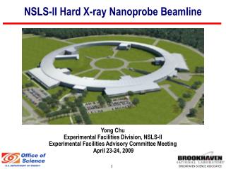
Hard x-ray nanoprobe beamline
10 likes | 181 Vues
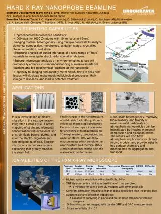
White beam slits. IVU20. Horizontal collimating mirror. Hard x-ray nanoprobe beamline. Beam Direction. Microscope Chamber. Detector Stand. ratchet wall. Horizontal Si(111) mono. Horizontal MLL. Detector Station.

Hard x-ray nanoprobe beamline
E N D
Presentation Transcript
White beam slits IVU20 Horizontal collimating mirror Hard x-ray nanoprobebeamline Beam Direction Microscope Chamber Detector Stand ratchet wall Horizontal Si(111) mono Horizontal MLL Detector Station Beamline Development Team:Yong S. Chu,Hanfei Yan, Evgueni Nazaretski, Jungdae Kim, Xiaojing Huang, Kenneth Lauer, Denis Kuhne Pink beam slits Horizontal focusing mirror Vertical MLL Storage Ring Mono beam slits HXN Satellite Building Beamline Advisory Team:I. C. Noyan (Columbia), D. Bilderback (Cornell), C. Jacobsen (ANL/Northwestern U.), A. Lanzirotti (U. Chicago), T. Buonasisi (MIT), S. Vogt (ANL), M. Holt (ANL), K. Evans-Lutterodt (BNL) Layers of IC Secondary source aperture 1 (Mature scope) CRL lenses for vertical focusing Diffraction Sample Stage Granite Block Multi-element XRF detector HXN Scientific CAPABILITIES Secondary source aperture 2 Separate Granite Block MLLs/ZP Area detector for CDI/DPC/Diffraction Sample • Unprecedented fluorescence sensitivity: • ~1000 cts/s for 1000 Zn atoms with 10nm focus at 10keV. • Imaging material heterogeneity using multiple contrasts to analyze elemental composition, morphology, oxidation states, crystalline phase, orientation, and strain. • Structural analysis of buried interfaces of a wide range of “hard” materials to investigate structure-functionality relations. • Spectro-microscopy analysis on environmental materials will dramatically enhance current understanding of mineral interfacial reactions and bio-geochemical reactions at the nanoscale. • Capability to imaging and quantify metal distributions in cells and tissues will elucidate metal-mediated biological processes, their linkage to diseases, and lead to potential treatment Applications Nucl. Instr. Meth. B. 199 (2003) after operation Aerosol Sci. Technol. 43 (2009) (La,Sr)(Co, Fe)O3 SOFC, Science, 325 (2009) Nano-scale heterogeneity, reactivity, bioavailability, and toxicity of environmental particulates or atmospheric nanoparticles can be investigated by imaging elemental composition and oxidation states. The results will also have atmospheric implications for climate forcing models, and provide insights into surface chemistry and mechanisms for aggregation and transport. In-situ investigation of electro-migration in the next-generation Integrated Circuits (IC). Parallel mapping of strain and elemental concentration will reveal evolution of strain fields before, during, and after the electro-migration and help mitigate its effects. Electron microscopy techniques require sectioning that greatly modifies the strain field. Small changes in the nanostructure of solid oxide fuel cells significantly influences macroscopic properties. Electron microscopy is inadequate for answering critical questions on 3D morphologies, composition, and oxidation states. HXN will allow measurements which correlate the nanostructure and chemical states at triple-phase boundaries with the macroscopic performance. Capabilities of the HXN X-ray microscope • Highest spatial resolution with scientific flexibility. • XRF fly-scan with a minimum dwell time of 1 ms per pixel. • 5 minutes for 5um x 5um 2D mapping with 10nm pixel size • Coherent diffraction imaging at higher spatial resolution than the probe size. • Substantial nano-diffraction capabilities • capable of analyzing in-plane and out-of-plane strain for crystalline • samples • Diffraction-contrast imaging with parallel XRF and DPC measurements • Strain sensitivity of ~10-4 MLL Module