Download
1 / 34
340 likes | 661 Vues
Osteomalacia related to vitamin D deficiency in a Patient with CVID. Case Report

E N D
Osteomalacia related to vitamin D deficiency in a Patient with CVID
Case Report 23 year-old woman diagnosed as having CVID in 1999 was admitted to the hospital by reason of evaluation of her general status in November of 2005. She did not have a complaint..
Medical History • Many hospitalizations caused by several recurrent airway tract infections and diarheae since the age of 11 years. • She was given a diagnosis of bronchiectasia in 1999.
Significanty low immunoglobulin levels • (IgG 225 mg/dl (700-1400) • IgM < 24 mg/dl (70-40) • IgA < 16 mg/dl (40-230) • Defective responses to vaccination with Haemophilus influenzae and tetanus toxoid, • Elimination of the causes related to secondary immunodeficiencies prompted the diagnosis of CVID and she was started on IVIG 400 mg/kg/month in 1999.
Medical history (2) • Lobectomy operation was performed in 2000. • She was also diagnosed with diabetes mellitus and has been undergoing insulin therapy since 2000. • Alaso diagnosed as having polyneuropathy and sensorineural hearing loss in 2003. • There was no history of consanguity. • 2-3 times/year hospitalization frequency while under therapy
Examination On Admission • Unremarkableapart from reduced sounds in the left lower part of the lung. • Blood pressure 110/80 mmHg • Temperature 36,5 centigrade • Pulse 90 beats/minute • 52 kg weight, BMI of 19,33
Laboratory On Admission Quantitative immunoglobulin (Ig) evaluation (Dade-Behring, nephelometry) revealed: IgG 660 mg/dl (700-1400) IgA and IgM undetectable.
The biochemical analyses revealed isolated high alkaline phosphatase (472 U/L, N. 90-260) levels. • Persistant finding since 1999 but no further evaluation had been performed.. • Other biochemical parameters including sedimentation rate (13 mm/h) and C-reactive protein(0,3 mg/L, N=0-5 mg/L)were all normal except for:
Albumin (g/dl) 4,1 3,5-5 • Globulin (g/dl) 2,2 2,5-3,5 • Vitamin B 12 (pg/ml) 243 197-866 • Folat (ng/ml) 8,16 3-16 • Vitamin A (mg/L) 1,7 0,3-1,1 • Beta-Carotene (micg/dl) 11,2 10-80 • Vitamin E (mg/L) 8,4 6-18 • Iron (micg/dl) 49 50-140 • Ferritin (mg/ml) 21,65 13-150 • Hct (%) 34,9 35-45 • HgbA1C (%) 6,0 < 6,5
Autoantibody profile including; Antinuclear Antimitochondrial Anti-smooth muscle Liver-kidney microsomal Gastric parietal cell Anti-neutrophil cytoplasmic antibodies Rheumatoid factor (RF) Anti-thyroid antibodies were negative.
The high resolution computed tomography (CT) of thorax and CT of the abdomen did not reveal granulomatous disease (liver size normal). • Endoscopy and biopsy revealed normal duodenal mucosa, there were no intraepithelial lymphocytes.
Osteocalcin was found to be increased (35,5-N: 3,1-13,7 ng/ml) • Deoxypyridinoline was normal (6,3 nM/mM N:3,0-7,4) • Parathyroid hormone (PTH) was normal (49 pg/mL N:15-65) • Urinary calciumwas low (25 mg/24 h N:100-300) • Calcium of 8,8 mg/dl (N:8,2-10,4) • Phosphate3,3 mg/dl (N:2,3-4,5) • Magnesium 2,2 mg/dl (N:1,5-2,6)
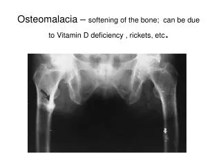
Radiology • The X-ray examination of the cranium and extremities revealed increased radiolucency without specific abnormalities suggestive of Paget’s disease. • The bone density evaluation showed osteoporosis. • The T score for the left hip was – 3.2 and for the lumbar spine was - 3.00. • The bone radionuclide scan was found to be normal.
25 OH vit D and 1,25 (OH) 2 vit D levels were found to be low. 25 OH vit D5 ng/ml (N: 7,6-75) 1,25 (OH) 2 vit D < 8 pg/ml (N:29,6-65,1) • Vitamin A 1,7 (N: 0,3-1,1 mg/L) • Vitamin E 8,4 (N: 6-18 mg/L)
Active vitamin D (rocaltrol 0,25 mcg bid and calcium (1 gr/day) therapy was started.
Rehospitalized 6 months later, by reason of breakdown of therapy.. • Osteocalcin normalized (11 ng/ml) • PTH increased (68 pg/ml) • ALP increased (508 U/L) • 25 OH vit D significantly low ( 1,2 ng/ml) • 1,25 OH vit D significantly low (1,2 pg/ml)
Active vitamin D (rocaltrol 0,5 mcg bıd and calcium therapy 1 gr/day were commenced.
ALP was normalized …….. 239 U/L (90-260) • Urinary calcium increased…..117 mg/24 h) • PTH was decreased no normal range……50,7 pg/ml (15-65) • 25 OH vit D was increased to………… 5 ng/ml (7,6-65) Still lower than normal…..
Possible Reasons for Low Vit D Levels • Living to the north of latitude 37 • Dark-skinned people, vegeterians and veiled women • Anticonvulsant or antituberculous drug use • Gastrointestinal and pancreatico-biliary diseases Celiac disease • Vitamin D resistance related to VDR mutation • Vitamin D binding protein deficiency were ruled out..
Vitamin D receptor expression (VDR) was evaluated and found to be significanly decreased in PBMC and hair follicle compared with control group…
VDR Gene Polymorphism She was found to be heterozygous regarding Apa, Bsm and Taq genes by High-Performance Liquid Chromatography (DHPLC).. She was homozygous (FF) for Fok gene..
None of these polymorphisms related to impaired VDR expression and function…
We could not investigate intestinal VDR expression • Low intestinal VDR expression is related to resistance to vit D treatment and calcium absorption…
Clinical Consequenses • Vitamin D deficiency might have a role in the pathogenesis of polyneuropathy in this patient. • Untreated vit D deficiency might play a role as an additive risk factor in terms of tendency to malignancy in patients with CVID. • Increased tendency to autoimmune diseases (type-1 diabetes !) • Increased tendency to infections (tuberculosis) • Increased tendency to sepsis • Irreperable bone loss and fractures
Isolation of total RNA and cDNA synthesis • 2 mL of blood and 10 follicles of hair sample of the case were collected. Fifty microliters of total RNA was isolated from peripheral blood mononuclear cells (PBMC) and hair follicle by using High Pure RNA Isolation Kit (Roche, Germany).Reverse transcription procedure was performed for cDNA synthesis by using Transcriptor First Strand cDNA Synthesis Kit according to the manufacturers’ instructions.
Relative quantification of VDR • Real-time quantitative RT-PCR analyses of VDR were performed with Lightcycler instrument and software. Glyceraldehyde-3-phosphate dehydrogenase (GAPDH “housekeeping” gene) was chosen as an internal standard to control for variability in amplification. The sequences of primers and probes used are shown in Table-3. PCR was performed by using TaqMan Master Kit (Roche Diagnostics) according to the instructions of the manufacturer. The VDR target probe was labeled at the 5’ end with the reporter dye molecule 6-carboxyfluorescein (FAM). The GAPDH target probe was labeled with 6- carboxyfluorescein. Both probes were labeled with the quencher fluor 6-carboxytetramethylrhodamine (TAMRA) at the 3’ end. To quantify VDR mRNA from PBMC and hair follicle, we constructed a calibration curve (Error: 0.100 Efficency: 1,790) using GAPDH mRNA as an endogenous control.