TRANSESOPHAGEAL ECHOCARDIOGRAPHY
2.41k likes | 11.56k Vues
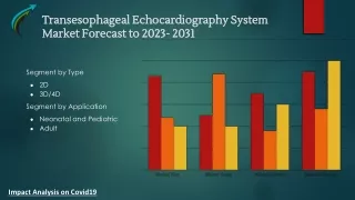
TRANSESOPHAGEAL ECHOCARDIOGRAPHY. Dr. Richa Jain. University College of Medical Science & GTB Hospital, Delhi. TRANSESOPHAGEAL ECHOCARDIOGRAPHY. Introduction Equipment Advantages Disadvantages Procedure Indications Contraindications Complications Clinical uses. INTRODUCTION.

TRANSESOPHAGEAL ECHOCARDIOGRAPHY
E N D
Presentation Transcript
TRANSESOPHAGEAL ECHOCARDIOGRAPHY Dr. Richa Jain University College of Medical Science & GTB Hospital, Delhi
TRANSESOPHAGEAL ECHOCARDIOGRAPHY • Introduction • Equipment • Advantages • Disadvantages • Procedure • Indications • Contraindications • Complications • Clinical uses
INTRODUCTION • In 1976, Dr Leon Frazin - concept of TEE. • Echocardiography:- the heart and great vessels probed with ultrasound (sound with frequency above 20 kHz). • Echocardiography uses ultrasound waves with frequency of 2.5 – 7.5 MHz. • Ultrasound sent into thoracic cavity and partially reflected by cardiac structures. • From these reflections: distance, velocity and density of objects within the chest derived.
INTRODUCTION ULTRASOUND WAVE AND ITS CHARACTERISTICS.
IMAGING TECHNIQUES • M MODE • One-dimensional views of cardiac structures produced by single-crystal transducers . • Density and position of all tissues in the path of a narrow ultrasound beam displayed as a scroll . • It is a timed motion display. • Principally used to view rapidly moving structures eg. valve leaflets. • Disadvantages: orientation and interpretation of spatial relationships difficult.
M-mode transesophageal echocardiogram of a normal aortic valve
IMAGING TECHNIQUES • 2D MODE • Rapid, repetitive scanning along many different radii within an area in the shape of a fan (sector). • A live (real time image) of heart is produced. • Advantage: the image obtained resembles an anatomic section and can be easily interpreted.
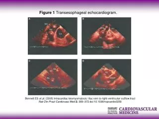
two-dimensional cross section of a normal aortic valve (AV)..
IMAGINGTECHNIQUES • DOPPLER TECHNIQUE:- • Based on doppler principle. • With doppler, blood flow velocity can be measured. • Different types of Doppler techniques: • Pulsed wave doppler • Continuous wave doppler • Colour flow doppler
Different types of doppler technique 1. Pulsed wave doppler:- • A small sampling volume (cursor) is placed in an area of interest with a 2D image. • Adv: measures blood flow velocities at selected areas of interest 3-5 mm wide along the ultrasound scan line. • Disadv: cannot measure fast blood flow velocities(>1m/s). • Use: to measure blood flow velocities through the pulmonary veins and mitral valve.
Pulsed wave Doppler echocardiogram of the main pulmonary artery (MPA).
Different types of doppler technique 2. Continuous wave doppler:- • Uses two sets of separate crystals: one to continuously emit ultrasound and one to continuously receive it. • Adv: detects blood flow velocities upto 7m/s. • Disadv: cannot identify location of the peak velocity • Use: to measure blood flow velocities through aorta, aortic valve,regurgitant valvular jets etc.
Different types of doppler technique 3. Colour flow doppler :- • Based on principle of PWD. • Uses multiple sample volumes along a scan line. • A colour code assigned to depict flow: toward (red) and away (blue) from the transducer. • 2 colour flow patterns • Normal aliasing pattern: due to laminar blood flow (as an area of homogenous color surface) • Mosaic pattern: due to turbulent blood flow ( as a mixture or mosaic of colour patterns known as colour jets)
Different types of doppler technique • “Normal” color Doppler aliasing • Mosaic pattern
Different types of dopplertechnique Colour doppler flow: • Adv: presents the spatial relationships between structure and blood flow. • Disadv: like PWD, it cannot measure fast blood flow velocities. • Use: to enhance recognition of valvular abnormalities, aortic dissections, and intracardiac shunts.
Different types of doppler technique TISSUE DOPPLER • A new use of PWD technology • To measure myocardial velocity. • It measures the velocity of the descent of the mitral annulus (Sm) towards the apex of the heart during normal LV contraction. • It decreases in presence of myocardial ischemia.
TEE EQUIPMENT • Monitor and TEE probe • TEE probe: a minaturizedechocardiographic transducer (40mm long, 13mm wide and 11 mm thick) mounted on the tip of a gastroscpoe. • Transducer: a phased array configuration with 64 piezoelectric elements operating at 3.7 to 7.5 MHz. • 2 knobs: one controls anteflexion and retroflexion; other controls rightward and leftward movement of the probe. • One electronic switch to scan the heart in various axial views .
DISADVANTAGES OF TEE • Semi invasive procedure : chances of injury • Needs special set up, technique, preparation, instrumentation • Needs orientation and expertise
PROCEDURE • Induction of anaesthesia and tracheal intubation • Patient’s neck extended • Well lubricated TEE probe introduced into the midline of hypopharynx with transducer facing anteriorly • Probe advanced into esophagus • During this manoeuvre, the control knob must be in neutral position.
Terminology used to describe transesophageal echocardiography probe movements.
I III II I- UPPER ESOPHAGEAL II- MID ESOPHAGEAL III- TRANSGASTRIC
Transesophageal echocardiography cross sections in a comprehensive examination.
INDICATIONS FOR PERIOPERATIVE TEE • Preoperative: hemodynamically unstable patients with suspected thoracic aortic aneurysms, dissection, or disruption • Intraoperative: • acute, persistent, and life-threatening hemodynamic disturbances • valve repair, CHD surgery for lesions requiring cardiopulmonary bypass; repair of hypertrophic obstructive cardiomyopathy; endocarditis; repair of aortic dissections; pericardial window procedures.
INDICATIONS FOR PERIOPERATIVE TEE • In ICU: unstable patients with unexplained hemodynamic disturbances, suspected valve disease, or thromboembolic problems.
CONTRAINDICATIONS OF TEE • ABSOLUTE: • Previous esophagectomy • Severe esophageal obstruction • Esophageal perforation • Ongoing esophageal haemorrhage • RELATIVE: • Esophageal diseases-diverticulum, varices, fistula • Previous esophageal surgery • Previous mediastinal irradiation • Unexplained swallowing difficulty
COMPLICATIONS OF TEE • Oral and pharyngeal injuries (0.1 – 0.3%) • Transient hoarseness (0.1 – 12%) • Esophageal injuries • Splenic injuries – 2 case reports • Endocarditis in outpatients
CLINICAL USES • EVALUATIONOFLVFILLING : • TEE reveals changes in left ventricular preload and filling pressure. • It measures EDA (end diastolic volume). EDA < 12cm2 - hypovolemia • Assessment of LV filling and function subjectively with the “trained eye”: a valid method to guide fluid administration. • Kuecherer : a systolic fraction of pulmonary venous flow < 55% - a sensitive and specific sign of LAP > 15mmH. ( as predominance of flow during diastole).
CLINICAL USES 2. ESTIMATION OF CARDIAC OUTPUT: • Real-time TEE images of LV filling and ejection permits qualitative, immediate detection of extreme changes in cardiac output. • TEE quantify CO the velocity and the cross-sectional area of blood flow. • SV = v x ET x CSA SV = stroke volume (ml) v = spatial average velocity of blood flow (cm/sec) ET = systolic ejection time (sec) CSA = cross-sectional area of the vessel (cm2 )
CLINICAL USES 3. Assessment of ventricular systolic function: • Fractional area change (FAC) during systole: a measure of global LV function. • FAC = EDA – ESA / EDA EDA : cross-sectional area at end diastole ESA : cross-sectional area at end systole. • Marked changes in FAC are apparent by simply viewing the real-time images. • Hallmarks of severe RV dysfunction: severe hypokinesis , enlargement of RV , change in shape of RV from crescent to round.
CLINICAL USES 4. Assessment of ventricular diastolic function: • TEE is an ideal tool for assessment of diastolic function because of its unobstructed view of the mitral valve and pulmonary veins. • Normal flow across the mitral valve in diastole has • E wave : an early higher-velocity component (generated by atrial pressure and ventricular relaxation) • A wave : lower-velocity component (generated by atrial contraction) • At slower heart rates, these two waves are separated by a period of relatively little flow (diastasis).
Line drawings representing simultaneous transesophageal pulsed wave Doppler recordings from the mitral annulus and right upper pulmonary vein.
CLINICAL USES 5. Detection of myocardial ischemia: • Acute myocardial ischaemia produce abnormal inward motion and thickening of affected myocardium. • Short axis view of LV at level of papillary muscle : best view • Wall thickening more specific marker than wall motion.
REFERENCES • Ronald .D. Miller: Transesophageal echocardiography. Miller’s Anaesthesia 7 th edition, 2010; 1329-1356. • Intraoperative echocardiography. Kaplan’s cardiac anaesthesia 3rd edition.


![[PDF READ ONLINE] Clinical Manual and Review of Transesophageal Echocardiography, 3/e](https://cdn7.slideserve.com/12522415/slide1-dt.jpg)