Download
1 / 52
530 likes | 661 Vues
Explore how ATCase and Hemoglobin are allosterically regulated, including enzyme subunits, quaternary structures, CTP inhibition, oxygen binding, and models for allosteric interactions.

E N D
Aspartate transcarbamolase is allosterically inhibited by the end product of its pathway Carbamoyl phosphate + aspartate N-carbamoylaspartate + Pi
Aspartate transcarbamolase • Catalyses the first step (the committed step) in the biosynthesis of pyrimidines (thiamine and cytosine), bases that are components of nucleic acids
Condensation of aspartate and carbomyl phosphate to form N-Carbamoylaspartate
How is the enzyme regulated to generate precisely the amount of CTP needed by the cell?
CTP inhibits ATCase, despite having little structural similarity to reactants or products
ATCase Consists of Separate Catalytic and Regulatory Subunits • Can be separated into regulatory and catalytic subunits by treatment with p-hydroxy-mercuribenzoate, which reacts with sulfhydryl groups
Mercurial dissociate ATCase into two subunits 11.6S PCMBS treated ACTase Native ACTase 2.8S 5.8S 2c3 + 3r2 c6r6 Ultracentrifugation Activity
Subunit characteristics • Regulatory subunit (r2) • Two chains (17kd each) • Binds CTP • No enzyme activity • Catalytic subunit (c3) • Three chains • Retains enzyme activity • No response to CTP
Structure of ATCase Cysteine binds Zn – PCMBS displaces Zn and destabilizes the domain
Use of PALA to locate active site Carbamoyl phosphate Aspartate Potent competitive inhibitor
The T-to-R state transition Each catalytic trimer has 3 substrate binding sites Enzyme has two quaternary forms.
CTP stabilises the T state • T state when CTP bound • Binding site for CTP in each regulatory domain • Binds 50Å from active site • allosteric
R and T state are in equilibrium Mechanism for CTP inhibition
ATCase displays sigmoidal kinetics Cooperativity R>T T>R
The importance of the changes in quaternary structure in determining the sigmoidal curve is illustrated by studies on the isolated catalytic trimer, freed by p-hydroxymercuribenzoate treatment. The catalytic subunit shows Michaelis-Menten kinetics with kinetic parameters indistinguishable from those deduced for the R-state. The term tense is apt – the regulatory dimers hold the two catalytic trimers close so key loops collide & interfere with the conformational adjustments necessary for high affinity binding & catalysis. Why does ATCase display sigmoidal kinetics
Basis for the sigmoidal curve(mixture of two Michaelis Menten enzymes) Low KM High KM
ATP is an allosteric activator R>T High purine mRNA synthesis ↑
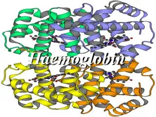
Myoglobin • Myoglobin is a single polypeptide, hemoglobin has four polypeptide chains. • Haemoglobin is a much more efficient oxygen-carrying protein. Why?
Myoglobin and Haemoglobin bind oxygen at iron atoms in heme 1 2 Fe2+ 3 4
Oxygen binding changes the position of the iron ion Sixth Co-ordination site Fifth Co-ordination site Proximal histidine
Quaternary structure of deoxyhemoglobin - HbA a1b1 and a2b2 dimers
Oxygen binding to myoglobin Simple equilibrium.
Haemoglobin as an allosteric protein • Haemoglobin consists of 2a and 2b chains • Each chain has an oxygen binding site, therefore haemoglobin can bind 4 molecules of oxygen in total • The oxygen-binding characteristics of haemoglobin show it to be allosteric
Oxygen binding to haemoglobin in rbc Cooperativity
Haemoglobin • Two principal models have been developed to explain how allosteric interactions give rise to sigmoidal binding curves • The concerted model • The sequential model
Concerted model • Oxygen can bind to either conformation, but as the number of sites with oxygen bound increases, so the equilibrium becomes biased towards one conformation (in the case of increasing oxygen bound, the R conformation)
Concerted model • Developed by Jacques Monod, Jeffries Wyman and Jeanne-Pierre Changeaux in 1965 • In this model all the polypeptide chains must be in an equilibrium that enables two possible conformations to exist
Concerted model • The concerted model assumes: • The protein interconverts between the two conformation T and R but all subunits must be in the same conformation • Ligands bind with low affinity to the T state and high affinity to the R state • Binding of each ligand increases the probability that all subunits in that protein molecule will be in the R state
Sequential model • Assumes • Each polypeptide chain can only adopt one of two conformations T and R. • Binding of ligand switches the conformation of only the subunit bound. • Conformational change in this subunit alters the binding affinity of a neighbouring subunit i.e. a T subunit in a TR pair has higher affinity that in a TT pair because the TR subunit interface is different from the TT subunit interface.
Sequential model • Devised by Dan Koshland in the 1950s • Substrate binds to one site and causes the polypeptide to change conformation • Substrate binding to the first site affects the binding of a second substrate to an adjoining site • And so on for other binding sites …
Quaternary structural changes on oxygen binding (T R) Rotation of a1b1 wrt a2b2 dimers
Haemoglobin must remain in T state in absence of oxygen T – state is extremely unstable
2,3-BPG (an allosteric effector) binds & stabilizes the T state (released in R state)
Fetal haemoglobin doesn’t bind 2,3-BPG so well so has higher oxygen affinity
Bohr effect (protons are also allosteric effectors) T-state stabilized by salt bridges Salt bridges Thus oxygen is released
Carbonic anhydrase Also … CO2 forms carbamate (R-NH-CO2) with N-ter – at interface between αβ dimers favours release of O2 by favouring the T state